Non ci sono recensioni
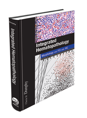
Unlike similar texts, which are often laden in technological details, Dr. Dunphy offers a comprehensive flow cytometry textbook, which covers in great depth, the technical aspects of FCI, phenotypic markers, as well as the advantages and disadvantages of FCI, but also a quick diagnostic guide incorporating normal and abnormal phenotypic findings of peripheral blood, body fluids, bone marrow, and lymph nodes. Integration of flow cytometry immunophenotyping with morphology and molecular genetic findings provides a complete diagnostic picture.
- The diagnostic applications of FCI in combination with morphology for all important hematolymphoid disorders.
- Detailed discussions of FCI mirror the new 2008 WHO classification, to increase the ease of use for practicing hematopathologists and residents.
- Separate chapters detail unique applications of FCI
- Brand new: 2008 WHO Classification
Cherie Dunphy, MD, is Executive Director of the Division of Hematopathology at the McLendon Clinical Laboratories and UNC Health Care in Chapel Hill and currently serves as president of the North Carolina Society of Pathologists. Integrated Hematopathology is the result of her decades of research on the application of flow cytometry, IHC, molecular and cytogenetic probes to diagnostic hematopathology. Her work has been documented in publications that include Archives of Pathology and Laboratory Medicine, AIMM (Applied Immunohistochemistry & Molecular Morphology), Acta Cytologica, Diagnostic Cytopathology, Cancer Genetics and Cytogenetic, and many other publications.
Table of Contents
xi Preface
1 General Introduction
1 Organization and Purpose
2 Applications of Flow Cytometry to
Diagnostic Hematopathology
2 References
2 Basic Principles and
Instrumentation of Flow
Cytometry
3 Basic Theory of FC
6 Antibodies and Fluorochromes
7 Sample Handling and Processing
9 References
3 Advantages and
Disadvantages of FCI
11 Specimen Requirements
12 Advantages of FCI
12 Disadvantages of FCI
15 FCI for Nodal and Extranodal
Tissues
15 FCI for BM Specimen Evaluation
16 FCI for Evaluation of
Lymphomatous Involvement of
Small Biopsies
16 References
4 Phenotypic Markers
Commonly Used by
FCI in Diagnostic
Hematopathology
19 Panhematopoietic Cell Antigens
19 HLA-DR (Immune-Associated)
Antigens
21 B-Cell Lineage-Associated
Antigens
24 Prognostic Markers in CLL
24 T-Cell Lineage-Associated
Antigens
26 Large Granular Lymphocyte- or
Natural Killer Cell-Associated
Antigens
27 Monocyte- and Myeloid-
Associated Antigens
30 Progenitor Cell-Associated
Antigens
31 Non-Lineage Antigens
36 Erythrocyte-Associated Antigens
36 Platelet-Associated Antigens
36 CD61
36 Paroxysmal Nocturnal
Hemoglobinuria (PNH)-Associated
Defi cient Antigens
37 Monoclonal Antibodies
as Targeted Therapy for
Hematolymphoid Malignancies
40 References
5 Normal vs Abnormal FCI
Findings: Peripheral Blood,
Body Fluids, Bone Marrow,
and Lymph Node
53 Introduction
54 Patterns of Light Scatter and
CD45 Expression
61 Patterns of Antigen Expression
65 Thymocytes, Thymoma, and
Blasts of Precursor T-Cell
Lymphoblastic Lymphoma/
Leukemia
72 References
FrontMatter.indd v 9/6/2009 9:30:19 PM
vi Integrated Hematopathology: Morphology and FCI with IHC
6 Classification of
Hematolymphoid Neoplasms
75 Chronic Myeloproliferative
Diseases (Myeloproliferative
Neoplasms, 2008)
75 Myelodysplastic/Myeloproliferative
Diseases
75 Myelodysplastic Syndromes
76 Acute Myeloid Leukemia (and
Related Precursor Neoplasms,
2008)
76 Precursor B-Cell Neoplasms
76 Precursor T-Cell Neoplasms
77 Mature B-Cell Neoplasms
77 Mature T-Cell and NK-Cell
Neoplasms
78 Hodgkin Lymphoma
78 Immunodefi ciency-Associated
Lymphoproliferative Disorders
78 Histiocytic and Dendritic Cell
Neoplasms
78 Mastocytosis
78 References
7 Myeloproliferative
Neoplasms
79 Classifi cation
79 Introduction
79 Chronic Myelogenous Leukemia
82 Non-CML MPNs
83 Summary
83 References
8 Myelodysplastic Syndromes
85 Classifi cation
85 Introduction
85 Diagnosis/Differentiation from
Various Benign Conditions
90 Grading of MDS
91 Predicting Prognosis, Leukemic
Transformation, and Relapse
92 Clues to Pathogenesis
92 Summary
93 References
9 Myelodysplastic/
Myeloproliferative Diseases
95 Classifi cation
95 Introduction
95 Chronic myelomonocytic leukemia
97 Juvenile Myelomonocytic
Leukemia
99 Comparison with Enzyme
Cytochemistry and
Immunohistochemistry
99 References
10 De-Novo Acute Myeloid
Leukemia
101 Classifi cation
101 Introduction
101 Diagnosis
107 Correlation of FCI With AML
Subtype
115 Detection of Minimal Residual
Disease and Relapse
115 FCI Compared With Enzyme
Cytochemical and IHC Techniques
119 References
11 Precursor B-Cell Neoplasms
123 Classifi cation
123 Introduction
123 Diagnosis
128 Detection of Minimal Residual
Disease and Relapse
128 Comparison of FCI vs IHC in
Precursor B-Lymphoblastic
Leukemia/Lymphoma
131 References
FrontMatter.indd vi 9/6/2009 9:30:19 PM
Integrated Hematopathology: Morphology and FCI with IHC vii
12 Mature B-Cell Neoplasms
133 Classifi cation
133 Introduction
136 B-Cell Chronic Lymphocytic
Leukemia/Small Lymphocytic
Lymphoma
144 B-Cell Prolymphocytic Leukemia
146 Mantle Cell Lymphoma
151 Follicular Lymphoma
154 Lymphoplasmacytic Lymphoma
157 Plasma Cell Neoplasms: Plasma
Cell Myeloma
161 Plasma Cell Neoplasms: Plasma
Cell Myeloma
162 Splenic Marginal Zone
B-Cell Lymphoma (± Villous
Lymphocytes)
168 Extranodal Marginal Zone B-Cell
Lymphoma of MALT Type
169 Nodal Marginal Zone B-Cell
Lymphoma (± Monocytoid B Cells)
170 Hairy Cell Leukemia
172 Diffuse Large B-Cell Lymphoma
179 Mediastinal (Thymic) Large B-Cell
Lymphoma
181 Intravascular Large B-Cell
Lymphoma
181 Primary Effusion Lymphoma
183 Burkitt Lymphoma/Burkitt Cell
Leukemia
185 Primary Cutaneous Marginal Zone
B-Cell Lymphoma
186 Primary Cutaneous Follicle Center
Lymphoma
187 Primary Cutaneous Diffuse Large
B-Cell Lymphoma, Leg Type
189 References
13 Precursor T-Cell Neoplasms
199 Classifi cation
199 Introduction
199 Diagnosis
206 FCI vs Immunohistochemistry
in Precursor T Lymphoblastic
Leukemia/Lymphoma
208 References
14 Mature T-Cell and Natural
Killer (NK)-Cell Neoplasms
209 Classifi cation
210 Introduction
215 Leukemic/Disseminated T-Cell
Prolymphocytic Leukemia (T-PLL)
216 T-Cell Large Granular Lymphocytic
Leukemia (LGLL)
220 Aggressive NK-Cell Leukemia
221 Systemic EBV+ T-Cell
Lymphoproliferative Disease of
Childhood
222 Adult T-Cell Leukemia/Lymphoma
(ATLL)
223 Primary Cutaneous Mycosis
Fungoides (Indolent)
224 Sézary Syndrome (Aggressive)
225 Primary Cutaneous CD30+ T-Cell
Lymphoproliferative Disorders
(Indolent)
225 Primary Cutaneous Anaplastic
Large Cell Lymphoma
227 Lymphomatoid Papulosis
227 Subcutaneous Panniculitis-Like
T-Cell Lymphoma (Indolent)
228 Hydroa Vacciniforme-Like
Lymphoma
228 Primary Cutaneous Small-Medium
CD4+ TCL (Indolent)
FrontMatter.indd vii 9/6/2009 9:30:19 PM
viii Integrated Hematopathology: Morphology and FCI with IHC
229 Primary Cutaneous Aggressive
Epidermotrophic CD8+ Cytotoxic
TCL
230 Primary Cutaneous γδ T-Cell
Lymphoma (Aggressive)
231 Extranodal NK/T-Cell Lymphoma,
Nasal Type (Aggressive)
231 Primary Cutaneous Peripheral
T-Cell Lymphoma, Unspecifi ed
(Aggressive)
231 Extranodal NK/T-Cell Lymphoma,
Nasal Type
233 Enteropathy-Type T-Cell
Lymphoma
237 Primary Extranodal Peripheral
T-Cell Lymphoma, Unspecifi ed
237 Nodal Angioimmunoblastic T-Cell
Lymphoma (AITL)
240 Peripheral T-Cell Lymphoma,
Unspecifi ed (PTCL-U)
241 Anaplastic Large Cell Lymphoma
245 CD4+CD56+ Hematodermic
Tumor, Alias “Blastic NK-Cell
Lymphoma”
247 References
15 Hodgkin Lymphoma
257 WHO Classifi cation
257 Introduction
257 Classical Hodgkin Lymphoma
261 Nodular Lymphocyte-Predominant
HL
264 References
16 Immunodeficiency-
Associated
Lymphoproliferative
Disorders
265 Classifi cation
265 Introduction
265 Lymphoproliferative Diseases
Associated with Primary Immune
Disorders
273 Lymphomas Associated with
Infection by HIV
276 Post-Transplant
Lymphoproliferative Disorders
278 Methotrexate (MTX)-Associated
Lymphoproliferative Disorders
278 References
17 Histiocytic and Dendritic
Cell Neoplasms
283 Classifi cation
283 Histiocytic Sarcoma
284 Langerhans Cell Histiocytosis and
Langerhans Cell Sarcoma
286 Interdigitating Dendritic Cell
Sarcoma (DCS), Follicular DCS,
and DCS, NOS
288 Disseminated Juvenile
Xanthogranuloma
288 Additional Applications of FC to
Dendritic Cells
288 References
FrontMatter.indd viii 9/6/2009 9:30:19 PM
Integrated Hematopathology: Morphology and FCI with IHC ix
18 Mastocytosis
289 Classifi cation
289 Introduction
289 Diagnosis
290 Immunophenotype of Neoplastic
Mast Cells
296 Differential Diagnosis
297 Mast Cell Sarcoma
297 Mast Cell Leukemia
298 References
19 FCI for Fine Needle Aspirate
Specimens
301 Recommended Triage Procedures
for FNAs
302 Initial Diagnosis of NHL
304 Evaluation of Recurrent NHL
304 Limitations of FNA Combined with
FCI in the Evaluation of Primary
and Recurrent Lymphomatous
Involvement
305 Classical Hodgkin Lymphoma
(cHL)
305 Composite Lymphoma
305 Situations Requiring Biopsy,
Based on FNA and FCI Results
305 Detecting Hematopoietic
Malignancy Granulocytic Sarcoma
Chloroma, Monocytic Sarcoma,
Erythroid Sarcoma
306 Determining Presence of
Metastatic Non-Hematolymphoid
Malignancy
306 References
20 FCI for Body Fluids
307 Introduction
307 Types of Specimens Suitable and
Specimen Requirements
307 Non-Hodgkin Lymphoma
310 Detecting Hematopoietic
Malignancy
312 FISH and PCR to Diagnose NHL
with Serous Effusions
312 Determining Presence of
Metastatic Non-Hematolymphoid
Malignancy
312 Limitations
312 References


