Non ci sono recensioni
- Presents tips and tricks for beginners and experts
- Provides educational material for 3D training courses
- Features comprehensively illustrated cases
- Includes an accompanying DVD with video clips of all sample cases
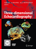
Three-dimensional echocardiography is the most recent fundamental advancement in echocardiography. Since real-time 3D echocardiography became commercially available in 2002, it has rapidly been accepted in echo labs worldwide.
This book covers all clinically relevant aspects of this fascinating new technology, including a comprehensive explanation of its basic principles, practical aspects of clinical application, and detailed descriptions of specific uses in the broad spectrum of clinically important heart disease.
The book was written by a group of well-recognized international experts in the field, who have not only been involved in the scientific and clinical evolution of 3D echocardiography since its inception but are also intensively involved in expert training courses. As a result, the clear focus of this book is on the practical application of 3D echocardiography in daily clinical routine with tips and tricks for both beginners and experts, accompanied by more than 150 case examples comprehensively illustrated in more than 800 images and more than 500 videos provided on a DVD.
In addition to an in-depth review of the most recent literature on real-time 3D echocardiography, this book represents an invaluable reference work for beginners and expert users of 3D echocardiography.
- Tips and tricks for beginners and experts
- Educational material for 3D training courses
- Comprehensively illustrated cases
- DVD with video clips of all sample cases
1 Introduction . . . . . . . . . . . . . . . . . . . . . . . . . . . 1
Thomas Buck, Andreas Franke,
and Mark J. Monaghan
2 Three-dimensional echocardiography:
lessons in overcoming time
and space . . . . . . . . . . . . . . . . . . . . . . . . . . . . . 3
Jos R.T.C. Roelandt and Joseph Kisslo
2.1 Reconstruction techniques . . . . . . . . . . . . . . . . . . 3
2.1.1 The linear scanning approach . . . . . . . . . . . . . . . 4
2.1.2 The fan-like scanning approach . . . . . . . . . . . . . 6
2.1.3 The rotational scanning approach . . . . . . . . . . . 6
2.2 Volumetric real-time (high-speed)
scanning . . . . . . . . . . . . . . . . . . . . . . . . . . . . . . . . . .10
2.3 The future is based on the past . . . . . . . . . . . . .17
References . . . . . . . . . . . . . . . . . . . . . . . . . . . . . . . . .18
3 Basic principles and practical
application . . . . . . . . . . . . . . . . . . . . . . . . . . . 21
Thomas Buck (with contribution by
Karl E. Thiele, Philips Medical Systems,
Andover, MA, USA)
3.1 How real-time 3D ultrasound works . . . . . . . .21
3.1.1 It’s all about the transmit beams . . . . . . . . . . . .21
3.1.2 The need for bigger, better, faster . . . . . . . . . .24
3.1.3 Fully sampled matrix array transducers . . . . .24
3.1.4 Parallel receive beam processing . . . . . . . . . . .26
3.1.5 Three-dimensional color flow . . . . . . . . . . . . . .27
3.2 The probes . . . . . . . . . . . . . . . . . . . . . . . . . . . . . . . .27
3.3 Live 3D echocardiographic
examination . . . . . . . . . . . . . . . . . . . . . . . . . . . . . . .29
3.3.1 First steps and learning curve . . . . . . . . . . . . . .29
3.3.2 Three-dimensional acquisition
(modes and image settings) . . . . . . . . . . . . . . . .29
3.3.3 Standard 3D views . . . . . . . . . . . . . . . . . . . . . . . . .34
3.4 Basic 3D analysis: cropping and
slicing . . . . . . . . . . . . . . . . . . . . . . . . . . . . . . . . . . . . .40
3.4.1 First steps of 3D dataset cropping . . . . . . . . . .40
3.5 Basic 3D measurements . . . . . . . . . . . . . . . . . . . .48
3.6 Artifacts . . . . . . . . . . . . . . . . . . . . . . . . . . . . . . . . . . .48
3.6.1 Stitching artifacts . . . . . . . . . . . . . . . . . . . . . . . . . .48
3.6.2 Dropout artifacts . . . . . . . . . . . . . . . . . . . . . . . . . .49
3.6.3 Blurring and blooming artifacts . . . . . . . . . . . .50
3.6.4 Gain artifacts . . . . . . . . . . . . . . . . . . . . . . . . . . . . . .51
References . . . . . . . . . . . . . . . . . . . . . . . . . . . . . . . . .53
4 Left ventricular function . . . . . . . . . . . . . . . 55
Harald P. Kühl
4.1 Assessment of left ventricular volumes
and ejection fraction . . . . . . . . . . . . . . . . . . . . . . .56
4.2 Evaluation of left ventricular mass . . . . . . . . . .59
4.3 Assessment of regional left ventricular
function . . . . . . . . . . . . . . . . . . . . . . . . . . . . . . . . . . .62
4.4 Determination of left ventricular
dyssynchrony . . . . . . . . . . . . . . . . . . . . . . . . . . . . . .65
4.5 Parametric imaging . . . . . . . . . . . . . . . . . . . . . . . .65
4.6 Transesophageal real-time 3D echocardiography
. . . . . . . . . . . . . . . . . . . . . . . . . . . . . .67
4.7 Three-dimensional assessment of left
atrial volume and function . . . . . . . . . . . . . . . . .68
References . . . . . . . . . . . . . . . . . . . . . . . . . . . . . . . . .70
5 Three-dimensional stress
echocardiography . . . . . . . . . . . . . . . . . . . . . 73
Andreas Franke
5.1 Method . . . . . . . . . . . . . . . . . . . . . . . . . . . . . . . . . . . .73
5.2 Clinical studies on 3D stress echocardiography
. . . . . . . . . . . . . . . . . . . . . . . . . . . . . .79
5.3 Limitations . . . . . . . . . . . . . . . . . . . . . . . . . . . . . . . .80
5.4 New approaches and future perspectives . . .80
References . . . . . . . . . . . . . . . . . . . . . . . . . . . . . . . . .80
6 Cardiac dyssynchrony . . . . . . . . . . . . . . . . . 83
Mark J. Monaghan and Shaumik Adhya
6.1 Technique . . . . . . . . . . . . . . . . . . . . . . . . . . . . . . . . .83
6.1.1 Acquisition of 3D volumes . . . . . . . . . . . . . . . . .83
6.1.2 Measures of intraventricular
dyssynchrony . . . . . . . . . . . . . . . . . . . . . . . . . . . . . .83
6.1.3 Performing 3D analyses . . . . . . . . . . . . . . . . . . . .86
6.1.4 Measurement variability . . . . . . . . . . . . . . . . . . .91
6.2 Normal values . . . . . . . . . . . . . . . . . . . . . . . . . . . . .92
6.3 Dyssynchrony in heart failure and left
bundle branch block . . . . . . . . . . . . . . . . . . . . . . .93
6.3.1 Relationship between left ventricular
function and dyssynchrony . . . . . . . . . . . . . . . .93
6.3.2 Patterns of dyssynchrony . . . . . . . . . . . . . . . . . . .95
6.3.3 SDI as a predictor of outcome of cardiac
resynchronisation therapy . . . . . . . . . . . . . . . . .95
6.4 Dyssynchrony due to right ventricular
pacing . . . . . . . . . . . . . . . . . . . . . . . . . . . . . . . . . . . . .96
6.5 Dyssynchrony in other situations . . . . . . . . . . .97
Table of contents
VIII Table of contents
6.5.1 In children . . . . . . . . . . . . . . . . . . . . . . . . . . . . . . . . .97
6.5.2 In congenital right heart disease . . . . . . . . . . .97
6.5.3 After acute myocardial infarction . . . . . . . . . . .97
6.5.4 In amyloidosis . . . . . . . . . . . . . . . . . . . . . . . . . . . . .98
6.6 Assessment of dyssynchrony after CRT . . . . .98
6.7 Conclusion . . . . . . . . . . . . . . . . . . . . . . . . . . . . . . . .98
References . . . . . . . . . . . . . . . . . . . . . . . . . . . . . . . . .99
7 The right ventricle . . . . . . . . . . . . . . . . . . . 101
Stephan von Bardeleben, Thomas Buck,
and Andreas Franke
7.1 Assessment of right ventricular volumes
and function . . . . . . . . . . . . . . . . . . . . . . . . . . . . . 104
7.2 New aspects of 3D right ventricular
analysis . . . . . . . . . . . . . . . . . . . . . . . . . . . . . . . . . . 107
References . . . . . . . . . . . . . . . . . . . . . . . . . . . . . . . 107
8 Valvular heart disease –
insufficiencies . . . . . . . . . . . . . . . . . . . . . . . 109
Thomas Buck
8.1 Mitral regurgitation . . . . . . . . . . . . . . . . . . . . . . 109
8.1.1 Evaluation of mitral valve insufficiency . . . 109
8.1.2 Classification of mitral valve
insufficiency . . . . . . . . . . . . . . . . . . . . . . . . . . . . . 111
8.1.3 Mitral valve prolapse, flail and Barlow’s
disease . . . . . . . . . . . . . . . . . . . . . . . . . . . . . . . . . . 112
8.1.4 Mitral valve quantification . . . . . . . . . . . . . . . . 116
8.1.5 Papillary muscle rupture . . . . . . . . . . . . . . . . . 121
8.1.6 Functional mitral regurgitation . . . . . . . . . . . 122
8.1.7 Endocarditis . . . . . . . . . . . . . . . . . . . . . . . . . . . . . 125
8.1.8 Mitral valve prosthesis . . . . . . . . . . . . . . . . . . . 126
8.1.9 Mitral valve repair . . . . . . . . . . . . . . . . . . . . . . . . 128
8.1.10 Rare etiologies . . . . . . . . . . . . . . . . . . . . . . . . . . . 132
8.1.11 Assessment of severity of mitral
regurgitation . . . . . . . . . . . . . . . . . . . . . . . . . . . . 133
8.2 Aortic regurgitation . . . . . . . . . . . . . . . . . . . . . . 143
8.3 Right-sided heart valves . . . . . . . . . . . . . . . . . . 149
References . . . . . . . . . . . . . . . . . . . . . . . . . . . . . . . 151
9 Valvular heart disease – stenoses . . . . . 155
Jose L. Zamorano and Jose Alberto de Agustín
9.1 Evaluation of mitral valve stenosis . . . . . . . . 155
9.1.1 Morphological assessment of the mitral
valve . . . . . . . . . . . . . . . . . . . . . . . . . . . . . . . . . . . . 155
9.1.2 Functional assessment of mitral stenosis . . 155
9.2 Evaluation of aortic valve stenosis . . . . . . . . 162
9.2.1 Morphological assessment of aortic
stenosis . . . . . . . . . . . . . . . . . . . . . . . . . . . . . . . . . 162
9.2.2 Functional assessment of aortic
stenosis . . . . . . . . . . . . . . . . . . . . . . . . . . . . . . . . . 164
9.3 Evaluation of tricuspid and pulmonary
valve stenosis . . . . . . . . . . . . . . . . . . . . . . . . . . . . 171
9.4 Evaluation of prosthetic and
reconstructed valves . . . . . . . . . . . . . . . . . . . . . . . 171
References . . . . . . . . . . . . . . . . . . . . . . . . . . . . . . . . . 172
10 Three-dimensional echocardiography
in adult congenital heart disease . . . . . . 175
Folkert J. Meijboom, Heleen van der Zwaan,
and Jackie McGhie
10.1 Patent foramen ovale . . . . . . . . . . . . . . . . . . . . . . 177
10.2 Atrial septal defect . . . . . . . . . . . . . . . . . . . . . . . . . 177
10.3 Ventricular septal defect . . . . . . . . . . . . . . . . . . . 182
10.4 Atrioventricular septal defects . . . . . . . . . . . . . 184
10.5 Ebstein’s anomaly . . . . . . . . . . . . . . . . . . . . . . . . . . 187
10.6 Transposition of the great arteries . . . . . . . . . 189
10.7 Congenitally corrected transposition
of the great arteries . . . . . . . . . . . . . . . . . . . . . . . . 191
10.8 Tetralogy of Fallot . . . . . . . . . . . . . . . . . . . . . . . . . . 193
10.9 RT3DE in other congenital cardiac
malformations . . . . . . . . . . . . . . . . . . . . . . . . . . . . . 193
10.10 The role of RT3DE in the analysis of
right ventricular function . . . . . . . . . . . . . . . . . . 193
10.10.1 Acquisition . . . . . . . . . . . . . . . . . . . . . . . . . . . . . . . . 193
10.10.2 Analysis . . . . . . . . . . . . . . . . . . . . . . . . . . . . . . . . . . . 196
10.11 Conclusion . . . . . . . . . . . . . . . . . . . . . . . . . . . . . . . . 196
References . . . . . . . . . . . . . . . . . . . . . . . . . . . . . . . . . 198
11 Congenital heart disease in
children . . . . . . . . . . . . . . . . . . . . . . . . . . . . . . 201
John M. Simpson
11.1 Technical and patient-specific factors . . . . . . 201
11.2 Selection of imaging probes in children . . . . 202
11.3 Presentation of 3D echocardiographic
images . . . . . . . . . . . . . . . . . . . . . . . . . . . . . . . . . . . . 202
11.4 Types of cardiac lesions which can be
assessed using 3D echocardiography . . . . . . 203
11.4.1 Abnormalities of venous drainage . . . . . . . . . 203
11.4.2 Atrial septal defects . . . . . . . . . . . . . . . . . . . . . . . . 203
11.4.3 Ventricular septal defects . . . . . . . . . . . . . . . . . . 204
11.4.4 Atrioventricular valves . . . . . . . . . . . . . . . . . . . . . 208
11.4.5 Atrioventricular septal defects . . . . . . . . . . . . . 208
11.4.6 Mitral valve abnormalities . . . . . . . . . . . . . . . . . 208
11.4.7 Ebstein’s anomaly of the tricuspid valve . . . . 208
11.4.8 Atrioventricular junction . . . . . . . . . . . . . . . . . . . 208
11.4.9 Complex anatomy . . . . . . . . . . . . . . . . . . . . . . . . . 212
11.5 Three-dimensional echocardiography
during catheter intervention and
surgery . . . . . . . . . . . . . . . . . . . . . . . . . . . . . . . . . . . . 214
11.5.1 Imaging during catheter intervention . . . . . . 214
11.5.2 Three-dimensional imaging during
surgery . . . . . . . . . . . . . . . . . . . . . . . . . . . . . . . . . . . . 214
11.6 Role of RT3DE in the assessment of
cardiac function in children . . . . . . . . . . . . . . . . 217
Table of contents IX
11.6.1 The left ventricle . . . . . . . . . . . . . . . . . . . . . . . . . . . 217
11.6.2 The right ventricle . . . . . . . . . . . . . . . . . . . . . . . 217
11.7 Conclusions . . . . . . . . . . . . . . . . . . . . . . . . . . . . . 219
References . . . . . . . . . . . . . . . . . . . . . . . . . . . . . . . 219
12 Cardiac tumors and sources of
embolism . . . . . . . . . . . . . . . . . . . . . . . . . . . . 223
Björn Plicht
12.1 Sources of embolism . . . . . . . . . . . . . . . . . . . . . 223
12.1.1 Cardiac and vascular thrombi,
spontaneous echo contrast . . . . . . . . . . . . . . 223
12.1.2 Patent foramen ovale . . . . . . . . . . . . . . . . . . . . 226
12.1.3 Infective endocarditis . . . . . . . . . . . . . . . . . . . . 231
12.2 Primary cardiac tumors . . . . . . . . . . . . . . . . . . 233
12.2.1 Primary benign cardiac tumors . . . . . . . . . . . 233
12.2.2 Primary malignant tumors . . . . . . . . . . . . . . . 236
12.3 Secondary cardiac tumors and
metastases . . . . . . . . . . . . . . . . . . . . . . . . . . . . . . 236
References . . . . . . . . . . . . . . . . . . . . . . . . . . . . . . . 239
13 Monitoring and guiding cardiac
interventions and surgery . . . . . . . . . . . . 241
Harald P. Kühl, Andreas Franke,
and Thomas Buck
13.1 Method . . . . . . . . . . . . . . . . . . . . . . . . . . . . . . . . . . 241
13.2 Intraoperative monitoring and guiding . . . 241
13.3 Periinterventional monitoring and
guiding . . . . . . . . . . . . . . . . . . . . . . . . . . . . . . . . . . 244
13.3.1 Transcatheter closure of PFO and ASD . . . . 244
13.3.2 Transcatheter closure of VSD . . . . . . . . . . . . . 248
13.3.3 Percutaneous aortic valve implantation . . . 248
13.3.4 Percutaneous mitral repair . . . . . . . . . . . . . . . 249
13.3.5 Left atrial appendage occluder
implantation . . . . . . . . . . . . . . . . . . . . . . . . . . . . . 256
13.3.6 Percutaneous occluder implantation
for paravalvular leaks . . . . . . . . . . . . . . . . . . . . 258
13.3.7 Percutaneous mitral valvuloplasty . . . . . . . . 259
13.3.8 Electrophysiological procedures . . . . . . . . . . 263
13.4 Conclusion . . . . . . . . . . . . . . . . . . . . . . . . . . . . . . 263
References . . . . . . . . . . . . . . . . . . . . . . . . . . . . . . . 263
Subject index . . . . . . . . . . . . . . . . . . . . . . . . 267


