Non ci sono recensioni
DA SCONTARE
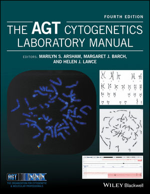
Cytogenetics is the study of chromosome morphology, structure, pathology, function, and behavior. The field has evolved to embrace molecular cytogenetic changes, now termed cytogenomics.
Cytogeneticists utilize an assortment of procedures to investigate the full complement of chromosomes and/or a targeted region within a specific chromosome in metaphase or interphase. Tools include routine analysis of G-banded chromosomes, specialized stains that address specific chromosomal structures, and molecular probes, such as fluorescence in situ hybridization (FISH) and chromosome microarray analysis, which employ a variety of methods to highlight a region as small as a single, specific genetic sequence under investigation.
The AGT Cytogenetics Laboratory Manual, Fourth Edition offers a comprehensive description of the diagnostic tests offered by the clinical laboratory and explains the science behind them. One of the most valuable assets is its rich compilation of laboratory-tested protocols currently being used in leading laboratories, along with practical advice for nearly every area of interest to cytogeneticists. In addition to covering essential topics that have been the backbone of cytogenetics for over 60 years, such as the basic components of a cell, use of a microscope, human tissue processing for cytogenetic analysis (prenatal, constitutional, and neoplastic), laboratory safety, and the mechanisms behind chromosome rearrangement and aneuploidy, this edition introduces new and expanded chapters by experts in the field. Some of these new topics include a unique collection of chromosome heteromorphisms; clinical examples of genomic imprinting; an example-driven overview of chromosomal microarray; mathematics specifically geared for the cytogeneticist; usage of ISCN’s cytogenetic language to describe chromosome changes; tips for laboratory management; examples of laboratory information systems; a collection of internet and library resources; and a special chapter on animal chromosomes for the research and zoo cytogeneticist. The range of topics is thus broad yet comprehensive, offering the student a resource that teaches the procedures performed in the cytogenetics laboratory environment, and the laboratory professional with a peer-reviewed reference that explores the basis of each of these procedures. This makes it a useful resource for researchers, clinicians, and lab professionals, as well as students in a university or medical school setting.
Table of Contents:
Contributing authors xxvii
Preface xxix
Acknowledgments xxxi
1 The cell and cell division 1
Margaret J. Barch and Helen J. Lawce
1.1 The cell 1
1.2 The cell cycle 14
1.3 Recombinant DNA techniques 19
1.4 The human genome 21
References 22
2 Cytogenetics: an overview 25
Helen J. Lawce and Michael G. Brown
2.1 Introduction 25
2.2 History of human cytogenetics 25
2.3 Cytogenetics methods 29
2.4 Slide‐making 49
2.5 Chromosome staining 58
2.6 Chromosome microscopy/analysis 59
2.7 Laboratory procedure manual 69
References 70
Contributed protocols 75
Protocol 2.1 Slide‐making 75
Protocol 2.2 Slide‐making 76
Protocol 2.3 Making wet slides for chromosome analysis 78
Protocol 2.4 Slide‐making 82
Protocol 2.5 Slide preparation 82
Protocol 2.6 Slide preparation procedure 84
3 Peripheral blood cytogenetic methods 87
Helen J. Lawce and Michael G. Brown
3.1 Using peripheral blood for cytogenetic analysis 87
3.2 Special uses of peripheral blood cultures 88
3.3 Peripheral blood constituents 89
3.4 Specimen handling 91
3.5 Cell culture equipment and supplies 93
3.6 Harvesting peripheral blood cultures 95
3.7 Chromosome analysis of peripheral blood 95
3.8 Storage of fixed specimens 95
Acknowledgments 95
References 95
Contributed protocols 98
Protocol 3.1 Blood culture and harvest procedure 98
Protocol 3.2 High‐resolution peripheral blood method 100
Protocol 3.3 Constitutional cytogenetic studies on peripheral blood 108
Protocol 3.4 Blood culture and harvest procedure for microarray confirmation studies 115
4 General cell culture principles and fibroblast culture 119
Debra F. Saxe, Kristin M. May and Jean H. Priest
4.1 Definitions of a culture 119
4.2 Basic considerations in cell culture 121
4.3 Fibroblast culture 128
4.4 Lymphoblastoid cell lines 132
Glossary 132
Reference 133
Additional readings 133
Contributed protocols section 134
Protocol 4.1 Solid tissue collection for establishing cultures 134
Protocol 4.2 Solid tissue transport and sendout media 135
Protocol 4.3 Tissue culture reagents 138
Protocol 4.4 Phosphate buffer solution deficient in Ca2+ and Mg2+ 141
Protocol 4.5 Solid tissue and fibroblast culture setup 141
Protocol 4.6 Solid tissue setup and processing 142
Protocol 4.7 Flask and coverslip setup for POC/fibroblast cultures 145
Protocol 4.8 Coverslip setup for solid tissue biopsy specimens 147
Protocol 4.9 Solid tissue (fibroblast) culturing and harvesting 150
Protocol 4.10 Fibroblast culture maintenance: media feeding and changing 154
Protocol 4.11 Routine subculture of fibroblast cultures 155
Protocol 4.12 Manual harvest for flasks 157
Protocol 4.13 Treated media for contamination 158
Protocol 4.14 Fungizone–mycostatin solution for treatment of fungus/yeast contaminated cultures 158
Protocol 4.15 Mycoplasma testing 159
Protocol 4.16 Plating efficiency of serum 160
Protocol 4.17 Routine replication plating for human diploid cells 160
Protocol 4.18 Cell counting chamber method 161
Protocol 4.19 Cell viability by dye exclusion 161
Protocol 4.20 Mitotic index 161
Protocol 4.21 Growth rate‐estimation of mean population doubling time during logarithmic growth 162
Protocol 4.22 Maintenance of fibroblast cultures as non‐mitotic population 163
Protocol 4.23 Synchronization at S‐phase with BrdU 163
Protocol 4.24 Making direct FISH preparations from abortus tissue 164
Protocol 4.25 Cryopreservation 165
Protocol 4.26 Cryopreservation with Nalgene cryogenic container 166
Protocol 4.27 Lymphoblastoid lines 167
Protocol 4.28 Freezing tissue cultures (cryopreservation) 171
5 Prenatal chromosome diagnosis 173
Kristin M. May, Debra F. Saxe and Jean H. Priest
5.1 Introduction 173
5.2 Amniotic fluid 173
5.3 Culture of amniotic fluid 175
5.4 Analysis of amniotic fluid 178
5.5 Chorionic villus sampling 180
5.6 Analysis of chorionic villi 184
References 186
Contributed protocols section 188
Protocol 5.1 Amniotic fluid culture setup and routine maintenance 188
Protocol 5.2 Coverslip (in situ) harvest procedure for chromosome preparations from amniotic fluid, CVS, or tissues (manual method) 191
Protocol 5.3 Harvest of flask amniocyte cultures 193
Protocol 5.4 Amniotic fluid culturing, subculturing, and harvesting (flask method) 195
Protocol 5.5 Criteria for interpreting mosaic amniotic fluid cultures 198
Protocol 5.6 Chorionic villi sampling – setup, direct harvest, and culture 199
Protocol 5.7 Chorionic villus sampling 204
Protocol 5.8 G‐Banding with Leishman’s stain (GTL) 208
Protocol 5.9 Cystic hygroma fluid protocol 209
6 Chromosome stains 213
Helen J. Lawce
6.1 Introduction 213
6.2 Chromosome banding methods 220
6.3 5-bromo-2′-deoxyuridine methodologies 246
6.4 T‐banding/CT‐banding 252
6.5 Antibody banding and restriction endonuclease banding 252
6.6 Destaining slides 252
6.7 FISH DAPI bands 252
6.8 Sequential staining 253
Acknowledgments 253
References 253
Contributed protocols section 266
Protocol 6.1 Conventional Giemsa staining (unbanded) 266
Protocol 6.2 Leishman’s stain 266
Protocol 6.3 Quinacrine mustard chromosome staining (Q‐bands) 266
Protocol 6.4 C‐banding 268
Protocol 6.5 C‐banding 270
Protocol 6.6 C‐banding 271
Protocol 6.7 C‐banding of blood slides 272
Protocol 6.8 Giemsa‐11 staining technique 274
Protocol 6.9 Distamycin A/DAPI staining 275
Protocol 6.10 Chromomycin/methyl green and chromomycin/distamycin fluorescent R‐banding method 277
Protocol 6.11 Bone marrow and cancer blood G‐banding 278
Protocol 6.12 Trypsin G‐banding 280
Protocol 6.13 Giemsa‐trypsin banding with Wright stain (GTW) for suspension culture slides and in situ culture coverslips 281
Protocol 6.14 G‐banding blood lymphocyte slides 284
Protocol 6.15 Cd staining 285
Protocol 6.16 CREST/CENP antibody staining 286
Protocol 6.17 AgNOR (silver staining) 287
Protocol 6.18 Sister chromatid exchange blood culture and staining 289
Protocol 6.19 Sister chromatid exchange fibroblast culture and staining 291
Protocol 6.20 T‐banding by thermal denaturation 294
Protocol 6.21 CT‐banding 295
Protocol 6.22 Lymphocyte culture and staining procedures for late replication analysis 295
Protocol 6.23 Destaining and sequential staining of slides 298
Protocol 6.24 Restaining permanently mounted slides 299
7 Human chromosomes: identification and variations 301
Helen J. Lawce and Luke Boyd
7.1 Understanding the basics 301
7.2 Description of human chromosome shapes 302
7.3 Determination of G‐banded chromosome resolution 355
Acknowledgments 356
Glossary 356
References 357
8 ISCN: the universal language of cytogenetics 359
Marilyn S. Arsham and Lisa G. Shaffer
8.1 Introduction 359
8.2 Language 359
8.3 Karyotype 364
8.4 Numerical events 378
8.5 Structural events 380
8.6 Derivative chromosomes (der) 394
8.7 Symbols of uncertainty 397
8.8 Random versus reportable 403
8.9 Multiple cell lines and clones 404
8.10 Fluorescence in situ hybridization 408
8.11 Microarray (arr) and region‐specific assay (rsa) 420
8.12 Conclusion 422
Acknowledgments 422
Addendum for ISCN 2016 updates 426
References 426
9 Constitutional chromosome abnormalities 429
Kathleen Kaiser‐Rogers
9.1 Numerical abnormalities 429
9.2 Structural rearrangements 444
References 472
10 Genomic imprinting 481
R. Ellen Magenis
10.1 Introduction 481
10.2 Human genomic disease and imprinting 488
10.3 Germ cell tumors – UPD and imprinting 493
Glossary 494
References 496
11 Cytogenetic analysis of hematologic malignant diseases 499
Nyla A. Heerema
11.1 Introduction 499
11.2 Myeloid leukemias 508
11.3 Myelodysplastic syndromes 514
11.4 Myeloproliferative neoplasms 515
11.5 B‐ and T‐cell lymphoid neoplasms 517
11.6 Lymphomas 522
11.7 Laboratory practices 525
Acknowledgments 533
Glossary of hematopoietic malignancies 533
References 535
Contributed protocols section 553
Protocol 11.1 Cancer cytogenetics procedure 553
Protocol 11.2 Bone marrow/leukemic peripheral blood setup and harvest procedure 558
Protocol 11.3 Bone marrow and leukemic blood culture and harvest procedure using DSP30 CPG oligonucleotide/interleukin‐2 for B‐cell mitogenic stimulation 560
Protocol 11.4 Culture of CpG‐stimulated peripheral blood and bone marrow in chronic lymphocytic leukemia 562
Protocol 11.5 Plasma cell separation and harvest procedure for FISH analysis 567
Protocol 11.6 Plasma cell separation and harvest procedure for FISH 569
Protocol 11.7 Bone marrow GTG‐banding 571
Protocol 11.8 GTW banding procedure (G‐bands by trypsin using Wright stain) 573
12 Cytogenetic methods and findings in human solid tumors 577
Marilu Nelson
12.1 Introduction 577
12.2 Processing tumor specimens 579
12.3 Recurrent cytogenetic abnormalities 592
12.4 Molecular genetic and cytogenetic techniques 608
12.5 Conclusion 612
Glossary 612
References 613
Contributed protocol section 631
Protocol 12.1 Solid tumor cell culture and harvest 631
Protocol 12.2 Solid tumor cell culture and harvest 637
Protocol 12.3 Solid tumor culture 643
Protocol 12.4 Solid tumor harvest: monolayer and flask methods 644
Protocol 12.5 Solid tumor culturing and harvesting 646
13 Chromosome instability syndromes 653
Yassmine Akkari
13.1 Introduction 653
13.2 Fanconi anemia 656
13.3 Bloom syndrome 658
13.4 Ataxia–telangiectasia 658
13.5 Nijmegen breakage syndrome 659
13.6 Immunodeficiency, centromeric instability, and facial anomalies syndrome 660
13.7 Roberts syndrome 661
13.8 Werner syndrome 661
13.9 Rothmund–Thomson syndrome 662
13.10 Proficiency testing 662
Glossary 662
References 667
Contributed protocol section 671
Protocol 13.1 Fanconi anemia chromosome breakage procedure for whole blood 671
Protocol 13.2 Supplemental procedure; Ficoll separation of whole blood 675
Protocol 13.3 Fanconi anemia fibroblast set up, culture, subculture, and harvest procedure 676
Protocol 13.4 Fanconi anemia chromosome breakage analysis policy 681
Protocol 13.5 Table for breakage studies result interpretation 682
Protocol 13.6 Fanconi anemia 684
14 Microscopy and imaging 687
Margaret J. Barch and Helen J. Lawce
14.1 The standard microscope 687
14.2 Brightfield microscopy 695
14.3 Fluorescence microscopy 697
14.4 Specialized microscopy 699
14.5 Capturing the microscopic image 701
References 703
15 Computer imaging 705
Christine E. Haessig
15.1 Introduction 705
15.2 Techniques to improve karyogram image quality 705
15.3 Metaphase preparation 706
15.4 Microscopy 706
15.5 Image capture 707
15.6 Enhancement 710
15.7 Advanced contrast 710
15.8 Macro programming 712
15.9 FISH imaging 713
15.10 Printing 715
15.11 Quality control 715
15.12 Archiving 715
Acknowledgments 715
References 715
16 Fluorescence in situ hybridization (FISH) 717
Helen J. Lawce and Jeffrey S. Sanford
16.1 Introduction 717
16.2 Clinical applications of FISH probes 722
16.3 Deletion/duplication probes for constitutional abnormalities 730
16.4 Hematology/oncology and solid tumor probes 734
16.5 Sources and characteristics of probes available to the clinical cytogenetics laboratory 736
16.6 Special uses of probes 738
16.7 Important FISH probe adjuvants 739
16.8 Principles of FISH 741
16.9 FISH methods – an overview 744
16.10 FISH analysis and reporting 757
16.11 FISH probe testing and validation 765
16.12 FISH for special investigation 768
16.13 Preimplantation genetic FISH 771
16.14 Other applications 776
16.15 Variants in FISH signal patterns 777
16.16 Conclusion 777
Acknowledgments 778
Glossary 778
References 780
Contributed protocols 790
Protocol 16.1 FISH (fluorescence in situ hybridization) methods 790
Protocol 16.2 LSI, CEP, and paint probe protocol 796
Protocol 16.3 FISH protocol for multiprobe® FISH panels 799
Protocol 16.4 Slide pretreatment with pepsin for FISH 800
Protocol 16.5 Interphase FISH for amniotic fluid specimen aneuploidy 801
Protocol 16.6 FISH on direct preparations from abortus tissue 803
Protocol 16.7 FISH on cultured non‐mitotic abortus tissue 804
Protocol 16.8 FISH on smears 806
Protocol 16.9 FISH on very small samples 808
Protocol 16.10 Paraffin‐embedded tissue FISH method 810
Protocol 16.11 VP2000 automated slide processor method for FFPE FISH 811
Protocol 16.12 Plasma cell targeted FISH 814
Protocol 16.13 Plasma cell separation for interphase FISH using easy SEP magnet method 815
Protocol 16.14 Preimplantation genetic testing (PGD) for aneuploidy 818
Protocol 16.15 Preimplantation genetic testing (PGD) FISH for translocations 821
Protocol 16.16 Post‐FISH BrdU antibody detection 823
Protocol 16.17 Same‐day HER2 IQ‐FISH pharmDx™ for breast tissue 824
17 Multicolor FISH (SKY and M‐FISH) and CGH 833
Turid Knutsen
17.1 Introduction 833
17.2 Multicolor FISH (SKY/M‐FISH) 834
17.3 Comparative genomic hybridization 849
17.4 Conclusion 859
Acknowledgments 859
References 859
Contributed protocols section 864
Protocol 17.1 Spectral karyotyping (SKY) 864
Protocol 17.2 Spectral karyotyping (SKY) 877
Protocol 17.3 DNA spectral karyotyping 878
Protocol 17.4 Multicolor‐FISH method (M‐FISH) I 881
Protocol 17.5 Multicolor FISH (M‐FISH) or 24‐color FISH II 884
Protocol 17.6 Multicolor FISH (M‐FISH) III 888
Protocol 17.7 Comparative genomic hybridization I 891
Protocol 17.8 Comparative genomic hybridization II 898
18 Genomic microarray technologies for the cytogenetics laboratory 903
Bhavana J. Davé and Warren G. Sanger
18.1 Introduction 903
18.2 Applications 907
18.3 Genomic microarray in a cytogenetics laboratory 913
18.4 Conclusion 922
Acknowledgment 922
Authors’ note 923
References 923
19 Mathematics for the cytogenetic technologist 937
Patricia K. Dowling
19.1 General concepts 937
19.2 Solutions 942
19.3 Statistical tools 956
19.4 Using a hemacytometer 968
19.5 Quantification and purity determination of DNA using spectroscopy 973
Reference 974
Additional readings 974
20 Selected topics on safety, equipment maintenance, and compliance for the cytogenetics laboratory 975
Helen Jenks and Janet Krueger
20.1 Introduction 975
20.2 Biological hazard safety 975
20.3 Chemical safety 980
20.4 Fire safety 986
20.5 Electrical safety 987
20.6 Disaster plan 988
20.7 Equipment operation, maintenance, and safety 988
20.8 Ergonomics 996
20.9 Regulatory considerations 998
Acknowledgments 1001
References 1001
Contributed protocols section 1003
Protocol 20.1 Autoclave sterilization, liquid nitrogen, pro‐par 1003
Protocol 20.2 Dishwashing procedure 1003
Protocol 20.3 Eppendorf pipette calibration 1004
Protocol 20.4 NIST thermometer calibration 1006
Protocol 20.5 Thermometer calibration 1008
Protocol 20.6 Timer calibration 1008
21 A system approach to quality 1011
Peggy J. Stupca and Sheryl A. Tran
21.1 Quality system 1011
21.2 Process management 1013
21.3 Documents and records 1015
21.5 Continual improvement 1022
21.6 Summary 1023
References 1023
Contributed protocols section 1025
Protocol 21.1 Quality control overview document 1025
Protocol 21.2 Monitoring specimen quality from off‐hill sites 1030
22 Laboratory management 1031
Mervat S. Ayad and Adam Sbeiti
22.1 Introduction 1031
22.2 Management concepts and functions 1032
22.3 Personnel management 1033
22.4 Quality management and control 1036
22.5 Budget development and monitoring 1039
22.6 Conclusion 1043
References 1043
Suggested reading 1043
23 Laboratory information system 1045
Peining Li and Richard Van Rheeden
23.1 Historical perspective 1045
23.2 General description of LIS 1045
23.3 LIS in cytogenetics laboratories 1048
23.4 Trends for the future LIS 1051
Acknowledgments 1052
References 1052
24 Animal cytogenetics 1055
Marlys L. Houck, Teri L. Lear and Suellen J. Charter
24.1 Introduction 1055
24.2 Domestic animal fertility 1056
24.3 Captive management 1057
24.4 Wildlife conservation 1059
24.5 General sample collection considerations 1060
24.6 Fibroblast cell culture 1062
24.7 Peripheral blood culture 1063
24.8 Chromosome analysis 1064
24.9 Molecular and comparative cytogenetics 1070
Acknowledgments 1071
Glossary 1072
References 1072
Contributed protocol section 1078
Protocol 24.1 Blood feather collection 1078
Protocol 24.2 Avian lymphocyte culture (for large birds) 1078
Protocol 24.3 Lymphocyte culture using whole blood 1084
Protocol 24.4 Lymphocyte culture using autologous plasma/buffy coat (AP/BC) 1085
Protocol 24.5 Horse lymphocyte culture method 1087
Protocol 24.6 Rhino blood culture 1089
Protocol 24.7 Organ tissue collection protocol from carcass 1090
Protocol 24.8 Skin biopsy procedure 1090
Protocol 24.9 Placenta biopsy procedure 1091
Protocol 24.10 Freezing of fibroblast cell cultures 1092
Protocol 24.11 Freezing tissue biopsy samples for later initiation of cell culture (tissue piecing) 1094
Protocol 24.12 Preparation of primary cultures from feather pulp 1095
Protocol 24.13 Preparation of primary cultures from solid tissue (explants) 1096
Protocol 24.14 Preparation of primary cultures using enzyme digestion 1097
Protocol 24.15 Harvesting of fibroblast cell cultures 1098
Protocol 24.16 Preparation of competitor DNA for FISH hybridization 1099
Protocol 24.17 In situ hybridization of BAC clones labeled with spectrum fluorochromes: probe and slide preparation 1100
Protocol 24.18 Labeling DNA with spectrum fluorochromes 1102
25 Online genetic resources and references 1103
Wahab A. Khan
25.1 Introduction 1103
25.2 Resource information 1103
Index 1113


