Non ci sono recensioni
DA SCONTARE
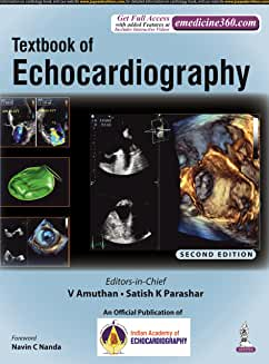
An echocardiogram uses sound waves to produce images of the heart. This common test allows a doctor to see the heart beating and pumping blood, and subsequently identify heart disease.
This book is a complete guide to performing and interpreting an echocardiogram. 56 chapters describe both basic and advanced techniques for diagnosing different heart disorders.
The second edition has been fully revised to provide clinicians with the latest developments and techniques in the field.
Seven new chapters have been added to this edition covering echocardiography and artificial intelligence, hypertension, arrhythmogenic right ventricular dysplasia, Kawasaki disease, cardio-oncology, diabetes mellitus, and foetal echo.
Dedicated chapters emphasise the role of echo in surgical procedures, and explore its use with electrophysiology – in patients with pacemakers and those undergoing cardiac resynchronisation therapy.
The book is highly illustrated with many 2D and 3D echo images helping explain the descriptive text for each topic.
CONTENTS
1. Basics of Ultrasound 1
Ashwin Venkateshvaran, Srikanth Sola
• Basic Principles 1; • Selecting a Transducer 2; • Understanding Image Resolution 3;
• Understanding Tissue Harmonic Imaging 5; • Understanding Artifacts 8
2. Introduction to Doppler Echocardiography 14
Madhu Mary Minz, Mansi Kaushik, Manish Bansal, Ravi R Kasliwal
• Doppler Equation 14; • Doppler Display 15; • Modes of Doppler Echocardiography 16;
• Doppler Ultrasound Equipment Control Panel 21; • Applications of Doppler Echocardiography in
Cardiology 22; • Doppler Artifacts 26
3. How to do Basic Transthoracic Echocardiography? 29
Vivek Tandon
• Preliminaries 29; • Basic Echocardiographic Examination 31; • Reporting 41
4. Performance of Transesophageal Echocardiography:
Basic Principles and Views 43
Nitin J Burkule, Manish Bansal
• Transesophageal Echocardiography Probe Hardware 43; • Patient Selection 43;
• Patient Set-up, Anesthesia, Intraprocedure Monitoring Considerations 44; • Technique of
Transesophageal Echocardiography Probe Insertion 46; • Technical Aspects of Transesophageal
Echocardiography Probe Manipulation and Image Display 47; • Transesophageal Echocardiography
Windows 49; • General Principles of Transesophageal Echocardiography Imaging 50;
• General Workflow of Comprehensive Transesophageal Echocardiography Imaging and Main Views 50
5. Chamber Quantification: Left Ventricle 65
Satish K Parashar, Rohit Tandon, Ankush Sachdeva, Sameer Shrivastava
• Measurement of Left Ventricular Linear Dimensions 66; • Left Ventricular Mass 67; • Evaluation of Left
Ventricular Volumes and Global Left Ventricular Systolic Functions 68; • Other Parameters for Estimating
Global Left Ventricular Functions 72; • Live Three-dimensional Echocardiography in Left Ventricular
Quantitation 75; • Global Longitudinal Strain 75 • Learning Points 77
6. Chamber Quantification: Right Ventricle 79
Rahul Mehrotra
• Problems with Right Ventricular Assessment 79; • Echocardiographic Views 79; • Measurement of
Right Ventricular Linear Dimensions 79; • Measurement of Right Ventricle Wall Thickness 82;
• Volumetric Assessment of Right Ventricle 82; • Measurement of Right Ventricle Systolic
Function 82; • Measurement of Right Ventricular Diastolic Function 84
Prelims.indd 21Prelims.indd 21 17-01-2022 17:55:1917-01-2022 17:55:19xxiiContents
7. Chamber Quantification: Left Atrium and Right Atrium 88
Rajan Joseph Manjuran
• Left Atrial Volume Assessment 88; • Right Atrial Volume Assessment 92
8. Echocardiography in Pulmonary Hypertension 95
R Alagesan, Satish C Govind
• Evolution of Criteria of Pulmonary Hypertension 95; • Definition and Classification 96;
• Echocardiography in Pulmonary Hypertension 96
9. Echocardiographic Evaluation of Diastolic Functions 111
Anirudddha De, Satish K Parashar
• What is Diastolic Dysfunction and Why LV Filling Pressure is Increased? 112; • Pathophysiology of
Diastolic Dysfunction 112; • Hemodynamic Concepts of Diastole 113; • Echocardiographic
Assessment of Diastolic Function 114; • Mitral Annular Tissue Doppler 115; • Additional Useful
Parameters 118; • Newer Diagnostic Parameters 122; • Grading of Diastolic Dysfunction 124;
• Assessment of Diastolic Dysfunction in Four Common Clinical Conditions 125; • Diastolic Stress Test 128
10. Echocardiographic Evaluation of Mitral Valve 132
Satish K Parashar, Rakesh Gupta, Nandita Chakrabarti, Sangeeta Porwal
• Anatomy of Mitral Valve 132; • Echocardiographic Evaluation of Mitral Valve 133
11. Echocardiographic Evaluation of Mitral Stenosis 137
Satish K Parashar, S Balasubramanian, I Balaji, V Amuthan, Rakesh Gupta
• Pathophysiology of Mitral Stenosis 137; • Etiology 138; • Diagnosis 140; • Stages of Mitral
Stenosis 141; • Assessment of Severity 141; • Grading of Severity 147; • Key Points in Valve
Morphology 148; • Intracardiac Thrombi in Mitral Stenosis 148; • Mimickers of Mitral Stenosis 150
12. Step-by-Step Evaluation of Mitral Regurgitation Using Echocardiography 154
Rohit Tandon, Satish K Parashar, Shibba Takkar, Gurpreet Singh Mahal
• Main Goals of Imaging 154; • Mitral Valve Anatomy 154; • Hemodynamic Effects of Chronic
Mitral Regurgitation 154; • Etiology of Mitral Regurgitation 154; • Mechanisms of Mitral
Regurgitation 156; • Advanced Echocardiography Techniques Used in Mitral Regurgitation 163;
• Role of Exercise Stress Echocardiography in Mitral Regurgitation Evaluation 164;
• Role of Echocardiography Imaging in Guiding Management Decisions 166
13. Examination of Valves: Aortic Valve 170
R Alagesan, Satish C Govind
• Anatomy of Aortic Valve 170; • Echocardiographic Examination 172
14. Echocardiography in Aortic Stenosis 179
R Alagesan, Satish C Govind
• Assessment of Aortic Stenosis 180; • Quantitative Doppler Echocardiography 184;
• Other Indices of Severity of Aortic Stenosis 195; • Correlation of Peak Gradient, Mean Gradient and
Effective Orifice Area 196; • Special Considerations in Aortic Stenosis 200
Prelims.indd 22Prelims.indd 22 17-01-2022 17:55:1917-01-2022 17:55:19Contentsxxiii
15. Echocardiography in Aortic Regurgitation 205
S Shanmugasundaram, U Ilayaraja, K Rajeswari
• Anatomy of Aortic Valve and Aortic Root 205; • Etiology of Aortic Regurgitation 206;
• Echocardiographic Evaluation 208
16. Echocardiographic Evaluation of the Aorta 227
Anezi Uzendu, Mahmoud Elsayed, Subash Dulal, Zeyad M Elmarzouky,
HK Chopra, Navin C Nanda
• Aortic Anatomy 227; • Two-dimensional Transthoracic Echocardiography 227; • Two-dimensional
Transesophageal Echocardiography 229; • Live/Real Time Three-dimensional Echocardiography 229;
• Aortic Aneurysm 230; • Aortic Dissection 230; • Aortic Intramural Hematoma 233;
• Aortic Transection 233; • Aortic Atheroma 233; • Aortic Tumor 233; • Marfan Syndrome 233
17. Echocardiography for Cardiac Prevention 236
Ankush Sachdeva, Sameer Shrivastava, HK Chopra
• Ankle–brachial Index 236; • Pulse Wave Velocity 237; • Carotid Intima-media Thickness 237;
• Left Ventricular Hypertrophy 238; • Left Atrial Volume Index 239; • Epicardial Fat 240;
• Aortic and Mitral Valve Calcification 241
18. Echocardiographic Assessment of Pulmonary Valve 244
G Rajesh
• Echocardiographic Imaging of the Normal Pulmonary Valve 244; • Echocardiographic Assessment
of Pulmonary Stenosis 247; • Echocardiographic Assessment of Pulmonary Regurgitation 249;
• Pulmonary Valve Assessment in Pulmonary Hypertension 250; • Pumonary Acceleration Time 250;
• Absent Pulmonary Valve 253; • Idiopathic Dilatation of Pulmonary Artery 253;
• Straight Back Syndrome 253
19. Echocardiographic Assessment of Tricuspid Valve 255
SR Veeramani, R Ramesh, V Thangamalar
• New Insights into Tricuspid Valve Anatomy and Physiology 255; • Imaging of the Tricuspid Valve 255;
• Imaging for Tricuspid Valve Intervention 256; • Tricuspid Valve Disease 256;
• Quantitative Measures of Tricuspid Regurgitation Severity 257; • Assessment of Tricuspid
Stenosis 260; • Evolving Concepts in Tricuspid Valve Intervention 260; • Transcatheter Tricuspid Valve
Intervention and Role of Imaging Modalities 260; • Future Directions 260
20. Prosthetic Heart Valve: Echocardiographic Evaluation 263
Vinayak Agarwal
• Historical Perspective 263; • Types of Valves 263; • Transcatheter Interventions 264;
• Two-dimensional Echocardiography 265; • Doppler in Evaluation of Prosthetic Heart Valve 271;
• Individual Prosthetic Heart Valve—Specific Evaluation 271; • Mitral Prosthetic Heart Valve 275;
• Special Features in Relation to PHV Evaluation 281; • Bioprosthetic Heart Valve Thrombosis 282;
• Special Considerations 283
21. Pulmonary Thromboembolism 288
Satish K Parashar, Rohit Tandon, S Balasubramanian, HK Chopra
• Pathophysiology and Predisposing Factors 288; • Severity of Acute Pulmonary Embolism Based on
Clinical Presentation 289; • Diagnosis of Pulmonary Thromboembolism 290; • Chronic Thromboembolic
Pulmonary Hypertension 293; • Serial Follow-up 294
Prelims.indd 23Prelims.indd 23 17-01-2022 17:55:2017-01-2022 17:55:20xxivContents
22. Tissue Doppler and Strain Imaging: Physical Principles and
Clinical Applications 298
Jagdish C Mohan, Madhu Shukla, Vishwas Mohan
• Mechanical Structure and Behavior of the Heart Muscle 299; • Myocardium: Architecture
and Function 300; • Myocardial Form and Function 301; • Descriptive Terms 303;
• Concept of Tissue Doppler Imaging 307; • Labeling of Tissue Velocity Waveforms 307;
• Technical Details of Tissue Doppler Imaging 309; • Myocardial Velocities in Short Axis 312;
• Internal Dependency of Velocities 314; • Clinical Applications of Tissue Doppler Imaging 317;
• How to Perform 2D Strain Imaging? 324; • Effects of Ischemia on Regional Deformation Metrics 326;
• Early Systolic Longitudinal Lengthening (Paradoxical Longitudinal Systolic Strain) 328;
• Postsystolic Longitudinal Shortening 329; • Paradoxical Strain Patterns 330; • Left Atrial Function
by Deformation Imaging 334; • Mechanical Properties of the Aorta 336; • 3D/4D Deformation
Imaging 336; • Strength and Weakness of Strain Imaging in Clinical Practice 339
23. Three-dimensional/Four-dimensional Echocardiography 344
V Amuthan, RVA Ananth
• Technical Development of Three-dimensional Echocardiography to Date 344;
• Procedure of Three-dimensional Echocardiography 345; • Cropping 348;
• Three-dimensional Transesophageal Echocardiography 348; • Four-dimensional Left Ventricular
Strain and Estimation Left Ventricular Torsional Deformation 351; • Three-dimensional Evaluation of
Left Ventricular Regional Function 353; • Three-dimensional Evaluation of Right Ventricular Function 356;
• Three-dimensional Evaluation of Mitral Valve 358; • Three-dimensional Evaluation of Mitral
Stenosis 360; • Three-dimensional Evaluation of Mitral Regurgitation 361;
• Three-dimensional Evaluation of the Aortic Valve 363; • Three-dimensional Evaluation of Aortic
Stenosis 367; • Three-dimensional Evaluation of Aortic Regurgitation 367;
• Tricuspid Valve 369; • Pulmonary Valve 369; • Three-dimensional Evaluation of Interatrial Septum,
Right And Left Atria 370; • Three-dimensional Evaluation of Left Atrium 371; • Three-dimensional
Evaluation of Left Atrial Appendage 371
24. Coronary Artery Disease 379
Manish Bansal, Ravi R Kasliwal
• Assessment of Regional Left Ventricular Systolic Function 379; • Diagnosis of Coronary Artery Disease
in the Outpatient Setting 381; • Diagnosis of Coronary Artery Disease in the Emergency Room 381;
• Assessing the Extent of Myocardial Damage 382; • Hemodynamic Assessment in Patients with
Myocardial Infarction 383; • Complications of Myocardial Infarction 383; • Detection of Residual
Myocardial Ischemia after Acute Management of Myocardial Infarction 386; • Prediction of Left Ventricular
Remodeling and Functional Recovery 387; • Risk of Ventricular Tachyarrhythmia and Sudden
Cardiac Death 389; • Other Applications of Echocardiography in Coronary Artery Disease 389
25. Stress Echocardiography for the Evaluation of Coronary Artery Disease 394
Nitin J Burkule, Manish Bansal
• Indications for Stress Echocardiography and Appropriate Usage Criteria 394; • Fundamental Principles
of Stress Echocardiography 396; • Setting Up a Stress Echocardiography Laboratory 396;
• Performance of Stress Echocardiography 397; • Diagnostic Accuracy of Stress Echocardiography 406;
• Role of Newer Modalities in Stress Echocardiography 408; • Prognostic Value of Stress
Echocardiography 410; • Additional Benefits of Stress Echocardiography 411;
• Clinical Relevance of Ischemia Detection with Stress Echocardiography 411; • Role of Stress
Echocardiography in Special Scenarios 412; • Versatility of Stress Echocardiography for
Improving its Diagnostic and Prognostic Value 412; • Assessment of Myocardial Viability 412;
Prelims.indd 24Prelims.indd 24 17-01-2022 17:55:2017-01-2022 17:55:20Contentsxxv
Appendix A 423
Appendix B 424
• Procedure 424; • ECG Findings 424; • Echocardiographic Findings 424; • Summary and
Interpretation 425; • Final Impression 425; • Procedure 426; • ECG Findings 426;
• Echocardiographic Findings 426; • Summary and Interpretation 427; • Final Impression 427
26. Myocardial Contrast Echocardiography 428
Nitin J Burkule, Manish Bansal
• Principles of Ultrasound Contrast Imaging 428; • Technique of Left Ventricular Cavity Opacification 428;
• Use of Left Ventricular Opacification in Clinical Practice 429; • Uses of Myocardial Contrast
Echocardiography in Clinical Practice 434; • Future Direction 435
27. Hypertrophic Cardiomyopathy 439
S Shanmugasundaram, B Vinodkumar, K Rajeswari
• Defining the Disease 439; • Inheritance 440; • Morphological Features 440;
• Hypertrophy: The Hallmark of HCM 440; • Myocardial Texture 446; • Crypts 446; • Ventricular
Function 446; • Left Atrial Abnormalities 449; • Mitral Apparatus 449; • Aorta and Aortic
Valve in HCM 450; • Altered Physiology 450; • Mitral Regurgitation in HCM 454; • Impaired Diastolic
Function 456; • Restrictive Phenotype 457; • LV Dyssynchrony 457; • Coronary Flow Reserve 457;
• Transesophageal Echocardiography 457; • 3D Echocardiography 458; • Stress
Echocardiography 458; • Contrast Echocardiography 458; • Role of Multimodality Imaging 458;
• Imaging to Differentiate HCM from Phenocopies 460; • Screening Family Members 462;
• Imaging to Guide Management Options 463; • Imaging for Risk Stratification 463;
• Follow-up Imaging 463
28. Echocardiography in Dilated Cardiomyopathy 467
CK Ponde
• Etiology 467; • Diagnostic Criteria for Dilated Cardiomyopathy Based on Imaging 467;
• Echocardiography Features in Dilated Cardiomyopathy 467; • Assessment of Systolic Functions in
Dilated Cardiomyopathy Using Conventional and New Techniques 469;
• Hemodynamic Assessment 471; • Dyssynchrony 472; • Prognosis 474
29. Restrictive Cardiomyopathy 479
Shantanu P Sengupta
• Types of Restrictive Cardiomyopathy 479; • Hemodynamics of Restrictive Physiology 479;
• Differentiation from Constrictive Pericarditis 479; • Cardiac Amyloidosis 481; • Sarcoidosis 482;
• Iron Overload: Hemosiderosis 483; • Eosinophilic Endomyocardial Disease 483; • Idiopathic (Primary)
Restrictive Cardiomyopathy 483
30. Role of Echocardiography in Diagnosis of Acute Rheumatic Fever 486
IB Vijayalakshmi
• Jones Criteria 486; • Echocardiography versus Clinical Examination 486; • Application of Vijaya’s
Echo Criteria 487; • Rheumatic versus Nonrheumatic Regurgitation 487; • Echo is the Key 488;
• Physiological versus Pathological Regurgitation 489; • Application of Revised Jones Criteria 2015 490
Prelims.indd 25Prelims.indd 25 17-01-2022 17:55:2017-01-2022 17:55:20xxviContents
31. Pericardium and Pericardial Diseases: An Echocardiographic Study 494
G Vijayaraghavan, Sakalesh Patil
• Congenitally Absent Pericardium 495; • Pericarditis 495; • Pericardial Effusion 496;
• Discollagenosis and Pericardium 498; • Uremic Pericarditis 498; • Cardiac Tamponade 499;
• Swinging Heart 502; • Ventricular Interdependence 503; • Low-pressure Tamponade—Cheshire Cat
Syndrome 504; • Loculated Effusions 504; • Effusive Constrictive Pericarditis 504;
• Constrictive Pericarditis 505; • Septal Bounce 506; • Annulus Paradoxus and Annulus
Reversus 507; • Transient Cardiac Constriction 507; • Constrictive Pericarditis and Restrictive
Cardiomyopathy 508
32. Pediatric Transthoracic Echocardiography: Basic Views 511
BRJ Kannan
• Sedation Protocol 511; • Basic Rules 511; • Echocardiographic Views 512
33. Echocardiographic Assessment of Acyanotic Congenital Heart Diseases 520
BRJ Kannan
• Classification of Acyanotic Congenital Heart Disease 520; • Chamber Dilatation in Atrial Septal
Defect 520; • Pulmonary Hypertension in Atrial Septal Defect 521;
• Ostium Secundum-Atrial Septal Defect 521; • Sinus Venosus-atrial Septal Defect 522;
• Ostium Primum-atrial Septal Defect 523; • Ventricular Septal Defects 523;
• Chamber Dilatation in Ventricular Septal Defect 523; • Pulmonary Hypertension versus Pulmonary Vascular
Disease in Ventricular Septal Defect 524; • Perimembranous Ventral Septal Defect 524;
• Outlet Ventricular Septal Defect 525; • Muscular Ventricular Septal Defect 526;
• Inlet Venricular Septal Defect 526; • Patent Ductus Arteriosus 527;
• Right Ventricular Outflow Tract Obstruction 528; • Subvalvular or Infundibular Stenosis 528;
• Valvular Pulmonary Stenosis 528; • Supravalvular Stenosis 528; • Left Ventricular Outflow Tract
Obstruction 528; • Valvular Aortic Stenosis 529; • Subvalvular Aortic Stenosis 529; • Supravalvular Aortic
Stenosis 529; • Coarctation of Aorta 529; • Assessment of Severity 530
34. Congenital Cyanotic Heart Disease 532
KM Krishnamoorthy, Deepa S Kumar
• Classification of Congenital Cyanotic Heart Disease 532; • General Aspects to Look for in All Patients
with CCHD 533; • Tetralogy of Fallot 535; • Pulmonary Atresia with Ventricular Septal Defect 537;
• Tricuspid Atresia 538; • Double Outlet Right Ventricle 538; • D-Transposition of Great
Arteries 539; • L-Transposition of Great Arteries 539; • Single Ventricle 540; • Hypoplastic Left Heart
Syndrome 540; • Pulmonary Atresia with Intact Ventricular Septum 541; • Ebstein’s Anomaly 542;
• Total Anomalous Pulmonary Venous Connection 544; • Truncus Arteriosus 545;
• Eisenmenger Syndrome 546; • Systemic Venous Anomalies Producing Cyanosis 547;
• Miscellaneous Conditions 547
35. Echo in the Emergency Room 552
Gurunath Parale, Chinmay Parale
• Cardiac Ultrasound in the Emergency Room: Focused versus Comprehensive Echo 552;
• Common Clinical Scenarios Where Echo is Required 552; • Evaluation of Chest Pain in the Emergency
Room 553; • Heart Failure 559; • Shock 561; • Cardiac Arrest 562;
• Stroke 565; • Chest Trauma 566; • Lung Ultrasound 566; • Medicolegal Issues 568
Prelims.indd 26Prelims.indd 26 17-01-2022 17:55:2017-01-2022 17:55:20Contentsxxvii
36. Cardiac Tumors 572
A George Koshy, Mathew Iype, KM Krishnamoorthy
• Clinical Presentations 572; • Benign Tumors 572; • Malignant Cardiac Tumors 577;
• Secondary Cardiac Tumors 579; • Normal Variants and Other Masses 579
37. Echocardiography in Infective Endocarditis 582
Chandramukhi Sunehra, P Krishnam Raju
• Incidence of Infective Endocarditis 582; • Epidemiology 582; • Diagnosis of Infective
Endocarditis 583; • Culture Negative Endocarditis 584; • Future Perspectives in the Molecular
Diagnosis of Infective Endocarditis 585; • Complications of Infective Endocarditis 588;
• Other Complications: Pericarditis, Myocarditis, and Myocardial Infarction 596; • Indications for Surgery
In Infective Endocarditis 596; • Checklist for Echocardiographic Assessment in a Patient with Infective
Endocarditis 598; • Echocardiography in Specific Clinical Situations of Infective Endocarditis 598;
• Right-sided Infective Endocarditis 599; • Fungal Endocarditis 600;
• Infective Endocarditis in Congenital Heart Disease 602; • Infective Endocarditis in Hypertrophic
Cardiomyopathy 605; • Limitations of Echocardiography in Infective Endocarditis 605;
• Newer Imaging Modalities 605
38. Cardiovascular Manifestations of Systemic Diseases 610
Chandramukhi Sunehra, P Krishnam Raju
• Systemic Inflammatory Diseases 610; • Granulomatous Disorders 616; • Metabolic Disorders 619;
• Thyroid Disorders 623; • Infiltrative Disorders 625; • Others Disorders 641; • Scorpion Stings and
Snake Bite 646; • Stress-induced Cardiomyopathy 646
39. Artifacts in Echocardiography 651
Sajan Ahmad Z, Anurag Bahekar
• Artifacts in Two-dimensional Echocardiography 651; • Artifacts in Spectral and Color Flow
Doppler 654; • Artifacts during Transesophageal Echocardiography 655; • Artifacts during
Contrast Echocardiography 656; • Artifacts in Three-dimensional Echocardiography 657
40. Interventional and Fusion Echocardiography 659
Sakshi Sachdeva, Anunay Gupta, S Ramakrishnan
• Interventional Echocardiography 659; • Fusion Echocardiography 660;
• Intracardiac Echocardiography 661
41. Echocardiography for Transcatheter Aortic Valve Replacement 664
Satish C Govind, R Alagesan
• Types of Valves 664; • Patient Selection 664; • Contraindications 665;
• Echocardiography 665; • Intraprocedural Echocardiography 672; • Complications 672;
• Postprocedure Echocardiography 674; • Advancements 675
42. Echocardiography in Percutaneous Mitral Valve Interventions 678
Harsha Basappa, Satish C Govind
• Techniques 678
Prelims.indd 27Prelims.indd 27 17-01-2022 17:55:2017-01-2022 17:55:20xxviiiContents
43. Intravascular Ultrasound 688
Vijayakumar Subban
• Principles and Imaging Systems 688; • Image Interpretation 688; • Clinical Applications of
Intravascular Ultrasound 689; • Clinical Evidence for Intravascular Ultrasound-guided Coronary
Intervention 694; • Intravascular Ultrasound in Specific Lesion Subsets 694;
• Future Perspectives 697
44. Echocardiographic Examination of a Patient with Pacemaker 702
Soumitra Kumar
• Right Ventricular Apical Pacing: What is Wrong with It? 702; • Echocardiography in Evaluation of Effects
of Right Ventricular Pacing on Left Ventricular Function 702; • Can We Prevent or Reverse Detrimental
Effects of Right Ventricular Apical Pacing? 704; • Echocardiography in the Evaluation of Tricuspid
Regurgitation Following Cardiac Pacing 704; • Echocardiography in the Evaluation of Mitral Regurgitation
Following Cardiac Pacing 706; • Echocardiography in the Diagnosis of Cardiac Perforation by Pacemaker
Leads 706; • Echocardiography in the Assessment of Pacemaker Lead Infection (Endocarditis)
and Thrombosis 706
45. Echocardiography in Cardiac Resynchronization Therapy 709
Ajit Thatchil, V Amuthan
• Role of Echocardiography in Patient Selection for Cardiac Resynchronization Therapy 709;
• Echocardiographic Techniques Used to Select Patients for CRT 713; • Speckle Tracking-based
Techniques 716; • Role of Echocardiography in Targeting Lead Placement for CRT 717;
• Role of Echocardiography in Optimizing CRT Programming Postimplant 719;
• Myocardial Work Based Techniques 721
46. Left Atrial Appendage (Anatomy, Physiology, Echocardiography,
Percutaneous Left Atrial Appendage Closure Assessment) 726
Daljeet Kaur Saggu, P Krishnam Raju
• Anatomy 726; • Physiology and Hemodynamics 727; • Echocardiographic Imaging of Left Atrial
Appendage 727; • Doppler Assessment 729; • Echocardiographic-guided Left Atrial
Appendage Closure 729
47. Echocardiography for Interventions in Congenital Heart Diseases:
Left-to-right Shunt Lesions 735
Kothandam Sivakumar
• Ventricular Septal Defects 735; • Atrial Septal Defects 755; • Patent Ductus Arteriosus 763;
• Aortopulmonary Window 768
48. Echocardiography in Operation Theater 774
Naman Sastri
• Important Role in Modern Cardiac Surgery 774; • Important Role in Modern Noncardiac
Surgery 774; • Echocardiographic Technique 774; • Important Preparation of Perioperative
Echocardiography 775; • Valvular Heart Disease in Operation Theater 775; • Assessment of the
Patient for Mitral Valve Repair 776; • Transesophageal Echocardiography during Major Aortic Surgery
and Reconstruction 784; • Left Ventricular Function and Wall Motion Abnormalities Intraoperatively 788;
• Pathology of Aorta and Role of Transesophageal Echocardiography in Operation Theater for Acute Aortic
Syndrome 788; • Errors and Artifacts in Assessment in OT with Ascending Aorta 789;
Prelims.indd 28Prelims.indd 28 17-01-2022 17:55:2017-01-2022 17:55:20Contentsxxix
• Epicardial Probe Preparation 790; • Management of Hemodynamic Status by TEE in Operation
Theater 791; • Diastolic Function Assessment by TEE 791; • Practicality of Diastolic Dysfunction in
Operating Room 792; • Strain during Intraoperative Period 793; • TEE in OT to Evaluate Donor and
Recipient Heart for Heart Transplant 795
49. Echocardiographic Practices in India: Legal Issues 798
KK Aggarwal, HK Chopra, K Thangamuthu
• Medicolegal Aspects of Echocardiography 798; • Difference of Opinion 802;
• Fetal Ultrasound/Echocardiography: Illustrative Case and Judgment 803
Appendix 1 805
Appendix 2 806
50. Artificial Intelligence and Echocardiography: Current Status
and Future Perspective 807
Karthik Seetharam, Partho P Sengupta
• Transcendence of Artificial Intelligence 807; • Types of Machine Learning 807; • Rise of Deep
Learning 807; • Importance of Machine Learning in Echocardiography 808; • Augmented Clinical
Inference 808; • Improved Accuracy 808; • Myocardial Function Assessment 808; • Identification
of Unique Phenotypes in Echocardiography 808; • Role of Machine Learning in Speckle Tracking
Echocardiography 810; • Role of Machine Learning in Big Data 811; • Contemporary Views on
Machine Learning 811
51. Status of Echocardiography in Hypertension 814
Rohit Tandon, Shivam Dutt, Gurpreet S Wander, Satish K Parashar
• Guidelines on Echocardiography Testing in Hypertension 814; • Role of Echo in Hypertension 814;
• Future Goals in Hypertension Imaging 823
52. Arrhythmogenic Right Ventricular Cardiomyopathy 826
Jesu Krupa
• Definition and Classification 826; • Epidemiology 826; • Pathology 826; • Genetics 827;
• Diagnostic Criteria 827; • Case Example 831; • Differential Diagnosis of Arrhythmogenic Right
Ventricular Cardiomyopathy 831; • Natural History 832; • Echocardiography for the Diagnosis of
Arrhythmogenic Right Ventricular Cardiomyopathy 833; • Arrhythmogenic Right Ventricular
Cardiomyopathy in the Pediatric and Adolescent Population 833; • Arrhythmias and Sudden
Cardiac Death Risk 835
53. Role of Two-dimensional Echo in Evaluation of Coronary Artery Disease
in Kawasaki Disease 837
Saji Philip
• Clinical Signs and Symptoms 837; • Myocardial Involvement In Kawasaki Disease 838;
• Coronary Aneurysms 839; • Imaging of Coronary Artery 841; • Common Locations of Aneurysms
in Patients with Kawasaki Disease 842; • Techniques in Coronary Artery Echocardiogram, Various
Positions, and Views 842; • Measurement of Coronary Artery 842; • Pitfalls in Evaluation of Coronary
Artery 842; • Recommended Views for the Coronary Artery Delineation 844;
• Reporting Format 847
Prelims.indd 29Prelims.indd 29 17-01-2022 17:55:2017-01-2022 17:55:20xxxContents
54. Role of Echocardiography in Cardio-oncology 849
Hardeep Kaur Grewal, Meera R
• Cancer and Cancer Therapy-related Cardiac Involvement 849; • Role of Echocardiography 850
55. Echocardiographic Evaluation of Diabetes Mellitus 864
V Amuthan, SR Veeramani, RVA Ananth
• Diabetic Cardiomyopathy 864; • Echocardiography 864; • Strain Imaging in Decision Making of
Diabetic Cardiomyopathy 866
56. Fetal Echocardiography 870
UP Singh
• Impact of Prenatal Diagnosis of Congenital Heart Disease 870; • Fetal Cardiovascular Hemodynamics
and Pathophysiology 871; • Fetal Position and Various Views 872; • Brief about Fetal Cardiac
Defects 873; • Diagnosis of Cardiac Arrhythmia on Fetal Echo 879; • Disorders Exclusive to
Fetal Heart 879; • Fetal Echo Report Format 879
Index
