Non ci sono recensioni
DA SCONTARE
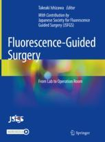
This volume is a practical guide of theranostics using intraoperative fluorescence imaging technology, as an all-out effort by the Japanese Society for Fluorescence Guided Surgery. It describes the various approaches the technique is being used such as vascular imaging, identification of lymphatic vessels by intratissue injection, lymph node imaging, and imaging for identification of anatomical structures. The book is organized into three major parts and the first one delivers the basics, introducing the use of the technology in clinical settings and initial setups. Next comes the description of clinical applications where chapters illustrate perfusion assessment, cancer localization, anatomy visualization, and lymph nodes/ducts mapping. Each chapter is devoted to the specific surgical field and disease areas, presenting images and videos of case studies. The last part presents some upcoming techniques for treatments. The Editor and the authors wish the ideas presented here will be hints to bridge the knowledge between surgeons and basic researchers for further innovation and practicality. It is important to stay up-to-date since intraoperative fluorescence imaging has been applied to clinical settings in various surgical fields and at the same time, novel techniques improving the efficacy of the technology have also been developed actively.
Fluorescence-Guided Surgery – From Lab to Operation Room is recommended for surgeons, operating nurses, medical experts, basic researchers and, industry engineers worldwide beyond boundaries of specialties. Edited and written by experts of The Japanese Society for Fluorescence-Guided Surgery, those who are the founders of the technology, it describes the accurate development history and cutting-edge techniques based on the knowledge accumulated over the years.
-
Front Matter
Pages i-xix
-
Part I
-
Front Matter
Pages 1-1
-
Clinically Available Fluorescent Reagents
- Kosuke Matsui, Masaki Kaibori
Pages 3-6
-
Indocyanine Green Fluorescence Imaging System for Open Surgery
- Nobuyuki Takemura, Norihiro Kokudo
Pages 7-12
-
Indocyanine Green Fluorescence Imaging System for Endoscopic and Robot-Assisted Surgeries
- Shuichi Watanabe, Toshiro Ogura, Hironobu Baba, Yusuke Kinugasa, Minoru Tanabe
Pages 13-17
-
5-Aminolevulinic Acid Fluorescence Imaging System
- Tsutomu Namikawa, Kazuhiro Hanazaki
Pages 19-23
-
How to Introduce Fluorescence Imaging to the Operating Room
- Sunao Uemura, Tsutomu Namikawa, Kazuhiro Hanazaki
Pages 25-27
-
Recording of Intraoperative Fluorescence Imaging
- Koshi Kumagai
Pages 29-31
-
-
Back Matter
Pages 33-36
-
Intraoperative Fluorescence Imaging [Practice] – Perfusion Assessment
-
Front Matter
Pages 37-37
-
- Masashi Yoshida
Pages 39-40
-
- Tohru Asai
Pages 41-45
-
Cerebral Angiography (Cerebral Aneurysm)
- Yasuo Murai, Fumihiro Matano, Akio Morita
Pages 47-54
-
Evaluation of Blood Perfusion in Skin Flaps
- Keisuke Okabe, Kazuo Kishi
Pages 55-62
-
Evaluation of Blood Perfusion in the Upper Gastrointestinal Tract
- Kazuo Koyanagi, Soji Ozawa, Yamato Ninomiya, Kentaro Yatabe, Itaru Higuchi, Miho Yamamoto
Pages 63-68
-
Evaluation of Blood Perfusion in Colorectal Surgery
- Hiro Hasegawa, Yuichiro Tsukada, Masaaki Ito
Pages 69-75
-
Perfusion Assessment in HBP Surgery and Liver Transplantation
- Satoru Seo
Pages 77-83
-
-
Back Matter
Pages 85-89
-
Part III
-
Front Matter
Pages 91-91
-
Liver Cancer (Primary Liver Cancer, Metastatic Liver Cancer)
- Yoshiharu Kono, Takeaki Ishizawa, Kiyoshi Hasegawa
Pages 93-99
-
Part III
-
Lung Cancer (Marking the Tumor Site)
- Toyofumi Fengshi Chen-Yoshikawa
Pages 101-110
-
Gastric Cancer (Primary Tumor, Peritoneal Dissemination)
- Tsuyoshi Takahashi, Yukinori Kurokawa, Makoto Yamasaki, Hidetoshi Eguchi, Yuichiro Doki
Pages 111-116
-
- Toshihiko Kuroiwa
Pages 117-125
-
- Keiji Inoue, Hideo Fukuhara, Shinkuro Yamamoto
Pages 127-133
-
-
Intraoperative Fluorescence Imaging [Practice] – Imaging of Lymph Nodes and Lymph Vessels
-
Front Matter
Pages 135-135
-
- Masashi Yoshida
Pages 137-137
-
Identification of Sentinel Lymph Nodes in Breast Cancer Surgery
- Manami Tada, Tomoharu Sugie
Pages 139-143
-
Identification of Sentinel Lymph Nodes in Gastric Cancer Surgery
- Shinichi Kinami
Pages 145-151
-
Identification of Sentinel Lymph Nodes in Colorectal Cancer Surgery
- Hironori Odaira, Masashi Yoshida, Yutaka Suzuki
Pages 153-157
-
Identification of Sentinel Lymph Nodes in Gynecologic Surgery
- Kensuke Sakai, Wataru Yamagami, Nobuyuki Susumu, Daisuke Aoki
Pages 159-164
-
Lymphography and Evaluation of Lymphedema
- Takumi Yamamoto
Pages 165-172
-
-
Part V
-
Front Matter
Pages 173-173
-
Imaging of the Bile Ducts (Fluorescence Cholangiography)
- Kazuhiro Matsuda, Takeshi Aoki, Tomokazu Kusano
Pages 175-182
-
- Takeshi Aoki, Kazuhiro Matsuda
Pages 183-193
-
- Yasuo Sekine
Pages 195-201
-
- Toshihiko Nishidate, Koichi Okuya, Kenji Okita, Ichiro Takemasa
Pages 203-210
-
Imaging of the Parathyroid Gland
- Akihiro Nakajo, Yoshiaki Shinden
Pages 211-216
-
-
Part VI
-
Front Matter
Pages 217-217
-
- Yasuteru Urano
Pages 219-229
-
Development of a New Imaging System
- Satoru Seo, Etsuro Hatano
Pages 231-236
-
Part VI
-
Development of a New Operating Room That Integrates Imaging Information
- Shunsuke Tsuzuki, Jun Okamoto, Manabu Tamura, Ken Masamune, Yoshihiro Muragaki
Pages 237-245
-
Therapeutic Applications: Photodynamic Therapy Using Porphyrin Compounds
- Takeomi Hamada, Atsushi Nanashima
Pages 247-251
-
Application to Therapy (2): Photoimmunotherapy Using a Near-Infrared Fluorescent Probe
- Yutaka Tamura, Akiko Suganami, Yoshiharu Okamoto
Pages 253-262
-
-
Back Matter
Pages 263-263
-
-


