Non ci sono recensioni
Dermatopathology is a subspecialty of both dermatology and pathology that studies causes and effects of skin disorders at a microscopic level.
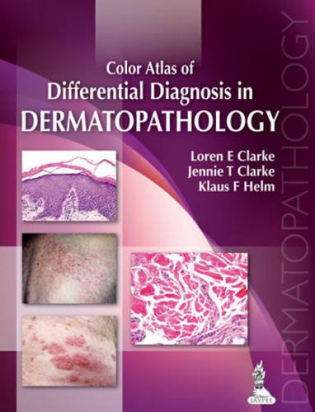
This colour atlas is a comprehensive guide to the diagnosis and management of dermatological disorders by correlating pathological results with clinical features. Written from the perspective of both pathologists and dermatologists, the book begins with an introduction to the normal pattern of skin.
Each of the following chapters examines a different skin disorder, first providing a detailed description of the disease and its clinical features, then criteria for diagnosis, differential diagnosis and potential pitfalls.
Written by an internationally recognised author team from Utah and Pennsylvania, this invaluable guide is highly illustrated with nearly 800 clinical and histological photographs with detailed descriptions.
Key points
- Comprehensive guide to diagnosis and management of skin diseases through pathology
- Highly useful for both dermatologists and pathologists
- Internationally recognised US author team
- Includes nearly 800 clinical and histological photographs
Author information
Loren E Clarke MD
Vice President of Medical Affairs, Dermatology Unit Myriad Genetics, Inc/MYGN, Salt Lake City, Utah, USA
Jennie T Clarke MD
Associate Professor of Dermatology
Klaus F Helm MD
Professor of Dermatology and Pathology
Both at Milton S Hershey Medical Centre, Penn State University, Hershey, Pennsylvania, USA
Contents
1.
Th
e Normal Skin Pattern
1
•
Findings within Stratum Corneum
3
•
F
indings within the Dermis
8
2.
Th
e Spongiotic and Psoriasiform Patterns
19
•
The Spongiotic Pattern
21
•
S
imulators of Spongiotic Dermatitis
29
•
The P
soriasiform Pattern
37
3.
Th
e Interface and Perivascular/Periadnexal Patterns
45
•
The Vacuolar Pattern
47
•
I
nterface Drug Eruption
55
•
The L
ichenoid Pattern
58
•
The Pit
yriasiform Pattern
70
•
The I
nterface and Perivascular/Periadnexal Patterns
72
4.
Th
e Blistering and Acantholytic Patterns
81
•
The Intraepidermal Blistering Pattern
85
•
The S
ubepidermal Blistering Pattern
103
5.
F
ollicular Processes
117
•
Infectious Bacterial Folliculitis
119
•
M
ajocchi’s Granuloma
120
•
H
erpes Zoster
121
•
N
oninfectious Causes of Folliculitis
122
•
Alop
ecia
125
6.
Th
e Nodular and Diffuse Dermal Infiltrative Patterns
133
•
The Granulomatous Pattern
135
•
The P
alisading Granulomatous Pattern
167
•
The N
eutrophilic/Suppurative Dermatitis Pattern
172
•
The Diffus
e Histiocytic Dermatitis Pattern
182
•
The L
ymphoplasmacytic Dermatitis Pattern
192
•
D
ermal Infestations and Arthropod Bite Reactions
194
7.
Th
e Vasculopathic Pattern
199
•
The Occlusive Vasculopathy Pattern
201
•
The A
cute Vasculitis Pattern
208
•
The F
ibrosing Vasculitis Pattern
216
•
V
asculitis with Macrophages/Granulomas
219
8.
Panniculitis
223
•
Septal Panniculitis
225
•
L
obular Panniculitis Lymphocytes Predominate
227
•
L
obular Panniculitis—Neutrophils Predominate
229
•
L
obular Panniculitis—Histiocytes Predominate
230
•
N
oninflammatory Panniculitides
232
9.
F
ibrosing Dermatitis
235
•
Diseases with Decreased Number of Fibroblasts
237
•
Dis
eases with Increased Number of Fibroblasts
241
Contents
xii
10.
Th
e Depositional Pattern
247
•
Deposits of Amorphous Eosinophilic Material (“Hyaline” Deposition)
249
•
Am
yloidoses
249
•
Mucinous/Myxoid Deposits
254
•
L
ichen Myxedematosus
254
•
Pr
etibial Myxedema (Localized Myxedema)
255
•
Other D
eposits
256
•
G
out (and Pseudogout)
256
•
C
alcinosis Cutis (and Osteoma Cutis)
257
11.
Th
e Melanocytic Tumors
259
•
Cytologically Banal Melanocytes Confined to the Epidermis
262
•
C
ytologically Atypical Melanocytes Confined to the Epidermis
268
•
C
ompound or Intradermal Neoplasms That are Cytologically Banal
272
•
C
ompound or Intradermal Neoplasms Composed of Spindled and/or Epithelioid Melanocytes:
Spitz’s and Reed’s Nevi
290
•
Pr
edominantly Dermal Tumors Composed of Pigment Synthesizing Melanocytes: The “Blue Nevus” Variants
293
•
C
ompound or Intradermal Neoplasms Composed of Markedly Atypical Melanocytes
301
12.
E
pithelial Neoplasms
325
•
Non-Neoplastic Epidermal Proliferations
327
•
B
enign Keratinocytic Neoplasms
331
•
M
alignant Tumors Arising from Epidermis
340
13.
A
dnexal Neoplasms
347
•
Criteria for Sebaceous Differentiation
349
•
T
ypes of Sebaceous Neoplasms
349
•
F
ollicular Neoplasms
354
•
F
ollicular Neoplasms Differentiating towards Infundibulum and Isthmus
354
•
T
umor of Hair Matrix
360
•
T
umors of External Root Sheath
361
•
T
umors of Hair Germ
362
•
T
umors of Hair Mesenchyme
364
•
A
pocrine Neoplasms
366
•
Cr
iteria for Apocrine Differentiation
366
•
N
eoplasms Exhibiting Follicular-Sebaceous Apocrine Differentiation
366
•
T
umors Exhibiting both Eccrine and Apocrine Variants
368
•
P
urely Apocrine Neoplasms
371
•
E
ccrine Neoplasms
373
•
Cr
iteria for Eccrine Differentiation (Diagnosis of Exclusion)
373
•
E
ccrine Neoplasms
373
•
C
ysts
376
14.
Th
e Mesenchymal Tumors
381
•
Paucicellular or Patternless Tumors
383
•
The F
ascicular and Storiform Tumors
399
•
The M
yxoid Tumors
423
•
The E
pithelioid Tumors
434
•
The L
ipomatous Tumors
444
•
The V
ascular Tumors
451
•
The P
leomorphic Tumors
472
•
The H
istiocytoid Tumors
475
•
The R
ound Cell Tumors
479
15.
Th
e Cutaneous Hematolymphoid Neoplasms
481
•
The Epidermotropic/Adnexotropic Pattern
483
•
The D
ermal ± Subcutaneous Pattern
492
•
The S
ubcutaneous Pattern
509
Index
511


