Non ci sono recensioni
DA SCONTARE
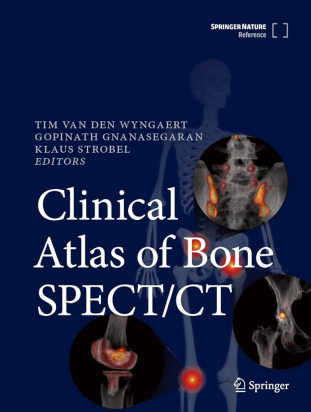
This clinical atlas is a comprehensive reference work on bone and joint disorders that can be characterized and assessed with hybrid bone SPECT/CT. It is structured according to the major joints and regions of the skeletal system, including spine, shoulder and elbow, hand and wrist, pelvis and hip, knee, and foot and ankle. For each region, the annotated normal X-ray and cross-sectional anatomy is presented, followed by a general introduction to the most common pathologies and frequent surgical procedures. Optimal bone SPECT/CT acquisition parameters are summarized and pre- and postoperative conditions are then discussed with the aid of informative clinical case vignettes featuring not only bone SPECT/CT images but also correlative findings on other imaging modalities. For every case, teaching points highlighting need-to-know findings and common pitfalls are presented. The book concludes with two dedicated chapters covering bone SPECT/CT imaging in sports injuries and oncology. Featuring many high-quality illustrations, Clinical Atlas of Bone SPECT/CT will be an invaluable resource for all nuclear medicine physicians.
It is published as part of the SpringerReference program, which delivers access to living editions constantly updated through a dynamic peer-review publishing process.
-
Front Matter
Pages i-xxiii
-
SPECT/CT Physics
-
Front Matter
Pages 1-1
-
Basic Radiation Physics: Radioactive Materials
- Tamar Willson
Pages 3-5
-
Basic Radiation Physics: X-rays
- Tamar Willson
Pages 7-9
-
- Tamar Willson
Pages 11-14
-
- Tamar Willson
Pages 15-18
-
-
SPECT/CT Technical Artifacts and Pitfalls
-
Front Matter
Pages 19-19
-
- Tamar Willson
Pages 21-24
-
- Tamar Willson
Pages 25-28
-
-
Spine
-
Front Matter
Pages 29-29
-
Normal Spine: X-ray and CT Anatomy
- Tim Van den Wyngaert
Pages 31-34
-
Spine: Introduction to Conditions and Procedures
- Tim Van den Wyngaert
Pages 35-38
-
Spine: Bone SPECT/CT Acquisition Protocol
- Tim Van den Wyngaert
Pages 39-42
-
Common Variants and Pitfalls: Transitional Vertebra
- Tim Van den Wyngaert
Pages 43-46
-
Common Variants and Pitfalls: Vertebral Hemangioma
- Tim Van den Wyngaert
Pages 47-53
-
Common Variants and Pitfalls: Bone Island (Enostosis)
- Tim Van den Wyngaert
Pages 55-59
-
Common Variants and Pitfalls: Limbus Vertebra
- Tim Van den Wyngaert
Pages 61-63
-
Common Variants and Pitfalls: Schmorl’s Node
- Tim Van den Wyngaert
Pages 65-69
-
Common Variants and Pitfalls: Scoliosis
- Tim Van den Wyngaert
Pages 71-73
-
Common Variants and Pitfalls: Fibrous Dysplasia
- Tim Van den Wyngaert
Pages 75-78
-
Common Variants and Pitfalls: Paget’s Disease
- Tim Van den Wyngaert, Klaus Strobel
Pages 79-82
-
Degenerative Spine: Bulging Disc
- Tim Van den Wyngaert
Pages 83-85
-
Degenerative Spine: Disc Herniation
- Tim Van den Wyngaert
Pages 87-90
-
Degenerative Spine: Degenerative Disc Disease
- Tim Van den Wyngaert
Pages 91-92
-
Degenerative Spine: Foraminal Stenosis
- Tim Van den Wyngaert
Pages 93-95
-
Degenerative Spine: Osteophytosis – Endplate Changes
- Tim Van den Wyngaert
Pages 97-100
-
Degenerative Spine: Facet Joint Arthropathy
- Tim Van den Wyngaert
Pages 101-104
-
Degenerative Spine: Spondylolysis
- Tim Van den Wyngaert
Pages 105-107
-
Degenerative Spine: Spondylolisthesis
- Tim Van den Wyngaert
Pages 109-112
-
Degenerative Spine Disease: Kyphosis
- Tim Van den Wyngaert
Pages 113-116
-
Degenerative Spine: Spinal Stenosis
- Tim Van den Wyngaert
Pages 117-119
-
Degenerative Spine Disease: Atlanto-occipital Degeneration
- Tim Van den Wyngaert
Pages 121-123
-
Degenerative Spine: Uncovertebral Joint Degeneration
- Tim Van den Wyngaert
Pages 125-127
-
Degenerative Spine: Vertebral Insufficiency Fracture
- Tim Van den Wyngaert
Pages 129-131
-
Inflammation and Infection: Baastrup’s Disease
- Tim Van den Wyngaert
Pages 133-136
-
Inflammation and Infection: Bertolotti’s Syndrome
- Tim Van den Wyngaert, Klaus Strobel
Pages 137-139
-
Inflammation and Infection: Spondylodiscitis
- Tim Van den Wyngaert
Pages 141-143
-
Inflammation and Infection: Diffuse Idiopathic Skeletal Hyperostosis (DISH)
- Tim Van den Wyngaert
Pages 145-147
-
Inflammation and Infection: Ankylosing Spondylitis
- Tim Van den Wyngaert, Klaus Strobel
Pages 149-153
-
Inflammation and Infection: SAPHO Syndrome
- Tim Van den Wyngaert
Pages 155-157
-
Inflammation and Infection: Scheuermann’s Disease
- Tim Van den Wyngaert
Pages 159-161
-
- Tim Van den Wyngaert
Pages 163-168
-
Bone Metastases (Lytic): Imaging Characteristics and Treatment Response
- Tim Van den Wyngaert
Pages 169-173
-
Bone Metastases (Sclerotic): Diagnostic Workflow and Imaging Characteristics
- Tim Van den Wyngaert
Pages 175-179
-
Malignant Vertebral Compression Fractures
- Tim Van den Wyngaert
Pages 181-184
-
- Tim Van den Wyngaert
Pages 185-188
-
- Tim Van den Wyngaert
Pages 189-192
-
- Tim Van den Wyngaert
Pages 193-196
-
Aneurysmal Bone Cysts: Imaging Characteristics
- Tim Van den Wyngaert
Pages 197-199
-
Postoperative Spine: Introduction
- Tim Van den Wyngaert
Pages 201-204
-
Postoperative Spine: Systematic Approach SPECT/CT Assessment
- Tim Van den Wyngaert
Pages 205-208
-
Postoperative Spine: Failed Back Surgery Syndrome (FBSS)
- Tim Van den Wyngaert
Pages 209-211
-
Postoperative Spine: Adjacent Segment Degeneration
- Tim Van den Wyngaert
Pages 213-216
-
Postoperative Spine: Hardware Failure – Pedicle Screw Loosening
- Tim Van den Wyngaert
Pages 217-219
-
Postoperative Spine: Cage Subsidence
- Tim Van den Wyngaert
Pages 221-223
-
Postoperative Spine: Pseudoarthrosis
- Tim Van den Wyngaert
Pages 225-228
-
Postoperative Spine: Locked Pseudoarthrosis
- Tim Van den Wyngaert
Pages 229-230
-
Postoperative Spine: Epidural Fibrosis
- Tim Van den Wyngaert
Pages 231-234
-
Postoperative Spine: Cervical Disc Arthroplasty
- Tim Van den Wyngaert
Pages 235-237
-
Pedicle Screw Fracture: Detection and Implications
- Tim Van den Wyngaert
Pages 239-240
-
Spine
-
Postoperative Spine: Dynamic Stabilization
- Tim Van den Wyngaert
Pages 241-243
-
-
Shoulder and Elbow
-
Front Matter
Pages 245-245
-
Normal X-ray and CT Anatomy of Shoulder and Elbow
- Biswajit Sahoo, Kanhaiyalal Agrawal
Pages 247-252
-
Bone SPECT/CT Acquisition Protocol in Shoulder and Elbow
- Girish Kumar Parida, Kanhaiyalal Agrawal
Pages 253-254
-
Introduction to Conditions and Procedures in Shoulder and Elbow
- Prateek Behera
Pages 255-257
-
Glenohumeral Osteoarthritis: An Overview
- Kanhaiyalal Agrawal, Girish Kumar Parida, Klaus Strobel
Pages 259-260
-
- Kanhaiyalal Agrawal, Girish Kumar Parida, Klaus Strobel
Pages 261-263
-
Osteoid-Osteoma of the Elbow: Imaging Findings
- Kanhaiyalal Agrawal, Girish Kumar Parida, Klaus Strobel
Pages 265-268
-
Heterotopic Ossification of the Elbow: Imaging Findings
- Kanhaiyalal Agrawal, Girish Kumar Parida, Klaus Strobel
Pages 269-271
-
Non-union or Delayed Union of the Elbow
- Kanhaiyalal Agrawal, Girish Kumar Parida, Klaus Strobel
Pages 273-275
-
- Kanhaiyalal Agrawal, Girish Kumar Parida, Klaus Strobel
Pages 277-282
-
Elbow Osteoarthritis: Imaging Findings
- Kanhaiyalal Agrawal, Girish Kumar Parida, Klaus Strobel
Pages 283-286
-
- Kanhaiyalal Agrawal, Girish Kumar Parida, Klaus Strobel
Pages 287-289
-
Insertion Tendinopathy of the Elbow: Imaging Findings
- Kanhaiyalal Agrawal, Girish Kumar Parida, Klaus Strobel
Pages 291-293
-
- Kanhaiyalal Agrawal, Girish Kumar Parida
Pages 295-297
-
-
Hand and Wrist
-
Front Matter
Pages 299-299
-
Hand and Wrist: Normal X-ray and CT Anatomy
- Ujwal Bhure, Klaus Strobel
Pages 301-302
-
Hand and Wrist: Introduction to Conditions and Procedures
- Ujwal Bhure, Klaus Strobel
Pages 303-303
-
Hand and Wrist: Bone SPECT/CT Acquisition Protocol
- Ujwal Bhure, Klaus Strobel
Pages 305-307
-
- Ujwal Bhure, Klaus Strobel
Pages 309-322
-
-
Hand and Wrist: Fracture Nonunion
- Ujwal Bhure, Klaus Strobel
Pages 323-332
-
Hand and Wrist: Carpal Boss and Painful Accessory Ossicles
- Ujwal Bhure, Klaus Strobel
Pages 333-339
-
Hand and Wrist: Kienbock’s Disease (Lunate Malacia)
- Ujwal Bhure, Klaus Strobel
Pages 341-347
-
- Ujwal Bhure, Klaus Strobel
Pages 349-356
-
Infections of the Hand and Wrist
- Ujwal Bhure, Klaus Strobel
Pages 357-360
-
Hand and Wrist: Complex Regional Pain Syndrome
- Ujwal Bhure, Klaus Strobel
Pages 361-366
-
Hand and Wrist: De Quervain Tenosynovitis
- Ujwal Bhure, Klaus Strobel
Pages 367-372
-
- Ujwal Bhure, Klaus Strobel
Pages 373-377
-
- Ujwal Bhure, Klaus Strobel
Pages 379-381
-
-
Pelvis and Hip
-
Front Matter
Pages 383-383
-
Hip and Pelvis: Introduction to Conditions and Procedures
- Arum Parthipun, Malavika Nathan
Pages 385-388
-
Hip and Pelvis: Bone SPECT/CT Acquisition Protocol
- Arum Parthipun, Malavika Nathan
Pages 389-390
-
Developmental Dysplasia of the Hip (DDH)
- Kanhaiyalal Agrawal, Komal Bishnoi, Geoffrey Chow, Thomas Armstrong, Tim Van den Wyngaert
Pages 391-397
-
- Kanhaiyalal Agrawal, Komal Bishnoi
Pages 399-401
-
- Kanhaiyalal Agrawal, Komal Bishnoi
Pages 403-406
-
Avulsion Fractures of the Hip and Pelvis
- Kanhaiyalal Agrawal, Komal Bishnoi, Geoffrey Chow, Thomas Armstrong
Pages 407-410
-
Arthritides: Osteoarthritis (OA)
- Kanhaiyalal Agrawal, Parneet Singh, Gopinath Gnanasegaran
Pages 411-414
-
Osteitis Pubis: Bone SPECT/CT Findings
- Tim Van den Wyngaert
Pages 415-418
-
- Kanhaiyalal Agrawal, Ujwal Bhure, Klaus Strobel
Pages 419-423
-
- Tim Van den Wyngaert
Pages 425-427
-
-
- Kanhaiyalal Agrawal, Tejasvini Singhal, Tim Van den Wyngaert
Pages 429-434
-
Femoroacetabular Impingement (FAI) Syndrome
- Kanhaiyalal Agrawal, Parneet Singh, Tim Van den Wyngaert
Pages 435-439
-
- Malavika Nathan, Arum Parthipun
Pages 441-443
-
Greater Trochanteric Pain Syndrome (GTPS) and Iliopsoas Bursitis
- Kanhaiyalal Agrawal, Tejasvini Singhal, Geoffrey Chow, Thomas Armstrong, Tim Van den Wyngaert
Pages 445-449
-
- Kanhaiyalal Agrawal, Ujwal Bhure, Geoffrey Chow, Thomas Armstrong, Klaus Strobel
Pages 451-456
-
Osteopoikilosis: Bone Scintigraphy and Imaging Features
- Kanhaiyalal Agrawal, Parneet Singh, Geoffrey Chow, Thomas Armstrong
Pages 457-461
-
Benign Bone Lesions: Sclerotic Enchondroma
- Kanhaiyalal Agrawal, Parneet Singh, Klaus Strobel
Pages 463-467
-
- Kanhaiyalal Agrawal, Ujwal Bhure, Klaus Strobel
Pages 469-472
-
Liposclerosing Myxofibrous Tumor of Femur
- Ujwal Bhure, Thomas F. Hany, Klaus Strobel
Pages 473-476
-
- Geoffrey Chow, Thomas Armstrong
Pages 477-479
-
- Geoffrey Chow, Thomas Armstrong
Pages 481-483
-
- Kanhaiyalal Agrawal, Ujwal Bhure, Klaus Strobel
Pages 485-490
-
Hip Replacement: Types of Arthroplasty and Fixation Zones
- Kanhaiyalal Agrawal, Tejasvini Singhal, Tim Van den Wyngaert
Pages 491-494
-
Hip Arthroplasty: Assessment of Component Position and Fixation
- Geoffrey Chow, Thomas Armstrong, Arum Parthipun, Malavika Nathan
Pages 495-499
-
Hip Arthroplasty: Normal Postoperative Findings
- Kanhaiyalal Agrawal, Tim Van den Wyngaert
Pages 501-506
-
Hip Arthroplasty: Abnormal Postoperative Findings
- Geoffrey Chow, Thomas Armstrong, Arum Parthipun, Malavika Nathan
Pages 507-514
-
Hip Arthroplasty: Periprosthetic Fractures
- Kanhaiyalal Agrawal, Tejasvini Singhal, Tim Van den Wyngaert, Klaus Strobel
Pages 515-519
-
Vascular Disorders: Legg-Calve-Perthes Disease
- Thomas Armstrong, Geoffrey Chow, Malavika Nathan
Pages 521-523
-
Avascular Necrosis of the Femoral Head
- Geoffrey Chow, Thomas Armstrong, Arum Parthipun, Malavika Nathan
Pages 525-527
-
-
Knee
-
Front Matter
Pages 529-529
-
-
Knee
-
Normal X-ray and CT Anatomy of the Knee
- Emma Robertson, Michael T. Hirschmann
Pages 531-532
-
Introduction to Knee Joint Conditions and Procedures
- Emma Robertson, Michael T. Hirschmann
Pages 533-535
-
- Edna Iordache, Michael T. Hirschmann
Pages 537-538
-
Osgood-Schlatter Disease: SPECT/CT Findings
- Emma Robertson, Helmut Rasch, Michael T. Hirschmann
Pages 539-541
-
Bone Bruise: SPECT/CT and MRI Findings
- Emma Robertson, Helmut Rasch, Michael T. Hirschmann
Pages 543-546
-
Ligament Pathologies: SPECT/CT, X-ray, and MRI Findings Pre- and Postoperative
- Emma Robertson, Helmut Rasch, Michael T. Hirschmann
Pages 547-552
-
Sinding-Larsen-Johansson Disease
- Emma Robertson, Helmut Rasch, Michael T. Hirschmann
Pages 553-555
-
Osteochondral Lesion of the Knee
- Emma Robertson, Helmut Rasch, Michael T. Hirschmann
Pages 557-560
-
Patellar Maltracking: Imaging Features
- Emma Robertson, Helmut Rasch, Michael T. Hirschmann
Pages 561-566
-
- Emma Robertson, Helmut Rasch, Michael T. Hirschmann
Pages 567-568
-
Osteochondritis Dissecans (OCD): Radiological Aspects and Treatment Options
- Edna Iordache, Helmut Rasch, Michael T. Hirschmann
Pages 569-572
-
Primary Osteoarthritis of the Knee: Radiological Aspects and Treatment Options
- Edna Iordache, Helmut Rasch, Michael T. Hirschmann
Pages 573-578
-
Post-traumatic Osteoarthritis (OA) of the Knee
- Edna Iordache, Helmut Rasch, Michael T. Hirschmann
Pages 579-582
-
Unicondylar Knee Arthroplasty (UKA): SPECT/CT Characteristics and Challenges
- Edna Iordache, Helmut Rasch, Michael T. Hirschmann
Pages 583-588
-
Patellofemoral Joint (PFJ) Arthroplasty
- Edna Iordache, Helmut Rasch, Michael T. Hirschmann
Pages 589-594
-
Painful Total Knee Replacement
- Edna Iordache, Helmut Rasch, Michael T. Hirschmann
Pages 595-604
-
-
Foot and Ankle
-
Front Matter
Pages 605-605
-
Foot and Ankle Pain: Introduction to Conditions and Procedures
- Dieter Berwouts, Laurent Goubau, Peter Burssens, Jeroen De Vil, Stefan Desmyter, Tom Lootens et al.
Pages 607-608
-
Foot and Ankle Bone SPECT/CT Acquisition Protocol
- Dieter Berwouts, Jeroen Mertens, Bieke Van Den Bossche, Bieke Lambert
Pages 609-611
-
- U. N. Pallavi, H. K. Mohan
Pages 613-619
-
-
- Patrick Martineau, Matthieu Pelletier-Galarneau
Pages 621-633
-
- Sherif Elsobky, Arum Parthipun
Pages 635-638
-
Osteoarthritis of the Foot and Ankle
- Dimitrios Priftakis
Pages 639-645
-
- Klaus Strobel, Ujwal Bhure, Thomas F. Hany, Tim Van den Wyngaert
Pages 647-652
-
Accessory Ossicles and Sesamoid Bones
- Dieter Berwouts, Laurent Goubau, Jeroen De Vil, Stefan Desmyter, Jeroen Mertens
Pages 653-665
-
Inflammatory and Infectious Conditions of the Foot and Ankle
- Dieter Berwouts, Laurent Goubau, Peter Burssens, Stefan Desmyter, Jeroen Mertens
Pages 667-673
-
- Dieter Berwouts, Tom Lootens, Jeroen De Vil, Jeroen Mertens
Pages 675-692
-
- Dieter Berwouts, Laurent Goubau, Tom Lootens, Jeroen De Vil, Jeroen Mertens
Pages 693-698
-
SPECT/CT in Osteochondroses and Osteonecrosis
- Dieter Berwouts, Laurent Goubau, Peter Burssens, Tom Lootens, Jeroen Mertens
Pages 699-704
-
Foot and Ankle: SPECT/CT Arthrography
- Dieter Berwouts, Stefan Desmyter, Peter Burssens, Jeroen Mertens
Pages 705-708
-
Recurrent Pain After Foot Arthrodesis: Diagnostic Value of Bone SPECT/CT
- Klaus Strobel, Ujwal Bhure, Tim Van den Wyngaert, Jeroen Mertens
Pages 709-713
-
- Dieter Berwouts, Stefan Desmyter, Tom Lootens, Jeroen Mertens
Pages 715-723
-
Forefoot Deformity Correction Surgery
- Dieter Berwouts, Jeroen De Vil, Peter Burssens, Jeroen Mertens
Pages 725-733
-
-
Sport Injuries
-
Front Matter
Pages 735-735
-
Epidemiology of Sporting Injuries
- Hans Van der Wall, Clayton Frater, Leticia Burton
Pages 737-743
-
Pathophysiology of Adult Sporting Injuries
- Hans Van der Wall
Pages 745-755
-
Pathophysiology of Pediatric Sporting Injuries
- Hans Van der Wall, John K. Pereira
Pages 757-765
-
Sporting Injuries in the Older Population
- Hans Van der Wall
Pages 767-773
-
Technical Factors in the Optimization of Scintigraphic Imaging
- D. W. Mackey, P. Chicco, L. Burton
Pages 775-781
-
Incidental Benign Skeletal Lesions: Bone Islands
- Gregory L. Falk, Scott B. Simpson
Pages 783-787
-
Incidental Benign Skeletal Lesions: Enchondroma
- Scott B. Simpson, Gregory L. Falk
Pages 789-791
-
Incidental Benign Skeletal Lesions: Fibrous Dysplasia
- Gregory L. Falk, Scott B. Simpson
Pages 793-795
-
Herniation Pit of the Femoral Neck
- Gregory L. Falk, Scott B. Simpson
Pages 797-800
-
Incidental Benign Skeletal Lesions: Non-ossifying Fibroma and Fibrous Cortical Defect
- Scott B. Simpson, Gregory L. Falk
Pages 801-804
-
Incidental Benign Skeletal Lesions: Osteochondroma
- Scott B. Simpson, Gregory L. Falk
Pages 805-808
-
Incidental Benign Skeletal Lesions: Osteoid Osteoma
- Gregory L. Falk, Scott B. Simpson
Pages 809-811
-
Incidental Benign Skeletal Lesions: Vertebral Hemangiomas
- Scott B. Simpson, Gregory L. Falk
Pages 813-815
-
Soft-Tissue Finding That May Lead to Confusion: Horseshoe Kidney
- Michael A. Magee, Leticia Burton, Hans Van der Wall
Pages 817-818
-
Soft-Tissue Finding That May Lead to Confusion: Polycystic Kidneys
- Michael A. Magee, Leticia Burton, Hans Van der Wall
Pages 819-820
-
Sporting Injuries in the Child and Adolescent
- John K. Pereira, Hans Van der Wall
Pages 821-832
-
- Saunders Jennifer, Robert Breit
Pages 833-839
-
Discitis: An Expected Cause of Low Back Pain in a Jogger
- Robert Breit, Hans Van der Wall
Pages 841-844
-
Pars Fractures of the Lumbar Spine
- Robert Breit, Jennifer Saunders, Hans Van der Wall
Pages 845-851
-
Occult Degenerative Disease in the Athletic Cohort
- Clayton Frater, Robert Breit, Hans Van der Wall
Pages 853-860
-
Septic Arthritis of the Acromioclavicular Joint
- Robert Breit, Hans Van der Wall
Pages 861-863
-
- Robert Breit, John K. Pereira, Hans Van der Wall
Pages 865-868
-
Acute and Chronic Elbow Injury
- Robert Breit, Hans Van der Wall
Pages 869-874
-
Multimodality Imaging of Shoulder Injuries
- Robert Breit, Hans Van der Wall
Pages 875-882
-
Injuries of the Wrist and Hand
- Hans Van der Wall, John K. Pereira, Clayton Frater
Pages 883-894
-
Central Low Back Pain and Pelvic Dysfunction
- Jennifer Saunders, Barbara Hungerford
Pages 895-903
-
-
Anterolateral Pelvic and Groin Pain
- Jennifer Saunders, Barbara Hungerford
Pages 905-911
-
Mechanical Dysfunction of the Sacroiliac Joint
- Jennifer Saunders, Barbara Hungerford
Pages 913-921
-
Functional Real Time Ultrasound in Optimal Muscle Activation in Disorders of the Sacroiliac Joint
- Barbara Hungerford, Jay Cunningham
Pages 923-927
-
Ancillary Changes Around the Trunk in Low Back Pain
- Jennifer Saunders, Barbara Hungerford
Pages 929-936
-
Thigh Splints: Adductor-Insertion Avulsion Syndrome
- Warwick J. M. Bruce, Siri Kannangara
Pages 937-939
-
- Warwick J. M. Bruce, Hans Van der Wall
Pages 941-946
-
Femoroacetabular Hip Impingement
- Warwick J. M. Bruce, Jennifer Saunders
Pages 947-951
-
Pathological Hip Fracture Due to an Aneurysmal Bone Cyst
- Warwick J. M. Bruce, Hans Van der Wall
Pages 953-954
-
- Warwick J. M. Bruce, Jennifer Saunders, Siri Kannangara
Pages 955-959
-
- Warwick J. M. Bruce, Michael A. Magee, Hans Van der Wall
Pages 961-970
-
Derangement of the Deep Knee Structures
- Warwick J. M. Bruce, Michael A. Magee, Hans Van der Wall
Pages 971-983
-
Medial Tibial Stress Syndrome (Shin Splints)
- Andrew Strokon, Hans Van der Wall
Pages 985-987
-
Stress Fracture of the Tibia and Fibula
- Andrew Strokon, Hans Van der Wall
Pages 989-994
-
- Andrew Strokon, Hans Van der Wall
Pages 995-1002
-
- Andrew Strokon, Hans Van der Wall
Pages 1003-1010
-
Ankle and Proximal Mid-Foot Pain
- Andrew Strokon, Hans Van der Wall
Pages 1011-1025
-
- Andrew Strokon, Hans Van der Wall, Clayton Frater
Pages 1027-1038
-
- Ujwal Bhure, Thomas F. Hany, Klaus Strobel
Pages 1039-1044
-
-
Bone Tumors/Oncology
-
Front Matter
Pages 1045-1045
-
Cross-Sectional Imaging in Skeletal Oncology
- Ankush Jajodia, Jitin Goyal
Pages 1047-1052
-
-
Bone SPECT/CT Acquisition Protocol in Skeletal Oncology
- Kanhaiyalal Agrawal
Pages 1053-1054
-
Benign Bone Tumors: Osteoid Osteoma
- P. Sai Sradha Patro, Kanhaiyalal Agrawal
Pages 1055-1058
-
Enchondroma and Chondroma: Benign Intramedullary Cartilage Tumors
- P. Sai Sradha Patro, Kanhaiyalal Agrawal, Klaus Strobel
Pages 1059-1062
-
Non-ossifying Fibroma, Fibrous Cortical Defect, and Fibroxanthomas: Benign Bone Tumors
- Kanhaiyalal Agrawal, P. Sai Sradha Patro
Pages 1063-1065
-
Benign Bone Tumors: Aneurysmal Bone Cyst
- P. Sai Sradha Patro, Kanhaiyalal Agrawal
Pages 1067-1069
-
Osteoblastoma: A Benign Bone Tumor
- Kanhaiyalal Agrawal, P. Sai Sradha Patro
Pages 1071-1073
-
Osteosarcoma: A Malignant Bone Tumor
- Kanhaiyalal Agrawal, P. Sai Sradha Patro
Pages 1075-1079
-
Chondrosarcoma: A Malignant Bone Tumor
- Kanhaiyalal Agrawal, P. Sai Sradha Patro
Pages 1081-1083
-
Malignant Bone Tumors: Ewing Sarcoma
- Kanhaiyalal Agrawal, P. Sai Sradha Patro
Pages 1085-1088
-
Giant Cell Tumors (GCT)/Osteoclastomas
- P. Sai Sradha Patro, Kanhaiyalal Agrawal
Pages 1089-1092
-
- Kanhaiyalal Agrawal, Gopinath Gnanasegaran
Pages 1093-1112
-
-
Skull and Jaw
-
Front Matter
Pages 1113-1113
-
Jaw: Normal X-ray and CT Anatomy
- Klaus Strobel, Ujwal Bhure
Pages 1115-1116
-
Jaw: Introduction to Conditions and Procedures
- Klaus Strobel, Ujwal Bhure
Pages 1117-1117
-
Jaw: Bone SPECT/CT Acquisition Protocol
- Klaus Strobel, Ujwal Bhure
Pages 1119-1120
-
SPECT/CT in Condylar Hyperplasia
- Klaus Strobel, Ujwal Bhure
Pages 1121-1126
-
SPECT/CT in Jaw Osteomyelitis and Osteonecrosis
- Klaus Strobel, Ujwal Bhure
Pages 1127-1133
-
- Klaus Strobel, Ujwal Bhure
Pages 1135-1136
-
Temporomandibular Joint Arthritis: Imaging
- Klaus Strobel, Ujwal Bhure
Pages 1137-1139
-
- Klaus Strobel, Ujwal Bhure
Pages 1141-1144
-
-
-
-
-
-
- Klaus Strobel, Ujwal Bhure
Pages 1145-1147
-
Skull: Normal X-ray and CT Anatomy
- Klaus Strobel, Ujwal Bhure
Pages 1149-1150
-
Skull: Introduction to Conditions and Procedures
- Klaus Strobel, Ujwal Bhure
Pages 1151-1151
-
Skull: Bone SPECT/CT Acquisition Protocol
- Klaus Strobel, Ujwal Bhure
Pages 1153-1154
-
Tumors and Tumor-Like Lesions in the Skull
- K. Strobel, U. Bhure
Pages 1155-1165
-
-
Anatomy
-
Front Matter
Pages 1167-1167
-
- Vanessa Quinn-Laurin, Ramin Mandegaran
Pages 1169-1173
-
- Vanessa Quinn-Laurin, Ramin Mandegaran
Pages 1175-1179
-
- Ramin Mandegaran, Vanessa Quinn-Laurin
Pages 1181-1185
-
- Vanessa Quinn-Laurin, Ramin Mandegaran
Pages 1187-1191
-
- Ramin Mandegaran, Vanessa Quinn-Laurin
Pages 1193-1197
-
- Ramin Mandegaran, Vanessa Quinn-Laurin
Pages 1199-1202
-
-
Back Matter
Pages 1203-1223
-
-
-
