Non ci sono recensioni
Features
-
- Reflects the latest technology in 3D echocardiographic imaging
- Presents top-quality illustrations demonstrating the latest advancements in diagnostic technology
- Demonstrates the clinical value of 3D echocardiography in catheter-based procedures, such as TAVI/TAVR and MitraClip
- Discusses the controversy of 3D echocardiography in cardiac resynchronization therapy
- Examines the evaluation of tricuspid valve morphology and function by transthoracic 3D echocardiography, including the forgotten tricuspid valve
Summary
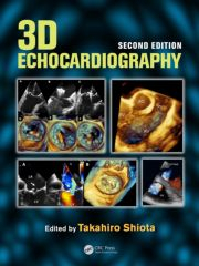
Since the publication of the first edition of this volume, 3D echocardiography has become a more frequent tool in diagnostic technology and patient care, while technology advancements have vastly improved this powerful imaging modality. Supplemented by video clips and illustrated with high-quality color images, 3D Echocardiography, Second Edition presents the work of experts in the field who disclose the latest findings and demonstrate the clinical value and advantages of modern 3D echocardiography over the traditional 2D imaging.
The book begins by describing the principles of 3D echocardiography and then proceeds to discuss its application to the imaging of:
- The left and right ventricle
- The left atrium
- Mitral stenosis and percutaneous mitral valvuloplasty
- Mitral regurgitation
- Aortic stenosis and regurgitation
- Tricuspid valve morphology
- Hypertrophic cardiomyopathy
- Congenital heart disease
The book also examines stress echocardiography and the use of 3D echocardiography in percutaneous valve procedures, cardiac resynchronization therapy, cardiac motion and deformation, and tissue tracking.
Table of Contents
Principles of 3D Echocardiographic Imaging; Bart Bijnens and Jan D’hooge
The Left Ventricle; Takeshi Hozumi and Junichi Yoshikawa
Stress Echocardiography; Andreas Franke
The Right Ventricle; Takahiro Shiota
The Left Atrium by 3D Echocardiography; Fabrice Bauer
Mitral Stenosis and Percutaneous Mitral Valvuloplasty; Jose Alberto de Agustín and Jose Zamorano
Primary (Organic) Mitral Regurgitation; Eric Brochet, Raluca Dulgheru, Giovanni La Canna, and Patrizio Lancellotti
Secondary Mitral Regurgitation; Patrizio Lancellotti, Raluca Dulgheru, Giovanni La Canna, Julien Magne, and Eric Brochet
Aortic Stenosis; Patrizio Lancellotti, Raluca Dulgheru, Julien Magne, and Eric Brochet
3D Echocardiography in the Evaluation of Aortic Regurgitation; Agnès Pasquet and Jean-Louis Vanoverschelde
Evaluation of Tricuspid Valve Morphology and Function by
Transthoracic 3D Echocardiography; Luigi P. Badano and Denisa Muraru
Functional Tricuspid Regurgitation; Jong-Min Song
Hypertrophic Cardiomyopathy; Marta Sitges, Carles Brambila, Takahiro Shiota, and Carles Paré
3D Echocardiography in Congenital Heart Disease; Philippe Acar
Aorta; Kyoko Otani, Masaaki Takeuchi, and Yutaka Otsuji
3D Echocardiography in Cardiac Resynchronization Therapy; Ulas Höke, Nina Ajmone Marsan, Jeroen J. Bax, and Victoria Delgado
3D Assessment of Cardiac Motion and Deformation; Nicolas Duchateau, Bart Bijnens, Jan D’hooge, and Marta Sitges
Tissue Tracking; Tomoko Ishizu, Shinichi Hashimoto, and Yoshihiro Seo
Index


